

The Numerous Alterations During Sample Preparation for EM Imaging
A huge problem for Virologists is that there is no way that they can claim what they present as evidence of a “virus” has any relation to natural particles that occur in vivo, or within a living organism. The various cell-altering chemicals/antibiotics and foreign animal RNA that is subsequently mixed together with the original unpurified sample (swab or BALF) from a sick individual immediately destroys any claim of purification (free of contaminants, pollutants, foreign materials). The fact that so much is added to the sample destroys the claim of isolation (separated from everything else).
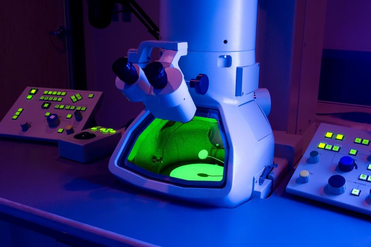
If the cell culturing process isn’t enough to alter the sample beyond it’s original state, the destruction of the natural cell morphology caused by preparing the sample for Transmission Electron Microscope images ensures that this is the case. Highlighted below are a few sources focusing on some of the steps (fixation and embedding) an already altered cell culture sample goes through in order to be prepared for imaging:
Preparation of cells for assessing ultrastructural localization of nanoparticles with transmission
electron microscopy
“To gain the resolution required to view individual NPs (<100 nm), electron microscopy is typically carried out. However, fragile biological samples such as cultured cells used in mechanistic studies require dehydration, heavy-metal staining and electron transparency for the sample to withstand the vacuum conditions and generate appropriate signal contrast to form an image. Further limitations of TEM include time consuming and toxic sample preparation, difficulties in distinguishing low-contrast nano-sized materials from cellular background features (e.g., cytoplasmic granules), production of two-dimensional black and white images and difficulty in drawing statistical conclusions. At present, stereological principles are being utilized to quantify the spatial distribution of immunogold and other NPs based on their localization throughout the cells and tissues for statistical evaluation25. These studies rely on χ2 analysis between treatment groups or within a single group to determine differences in uptake amount or localization to specific intracellular compartments. The purpose of these studies is being able to carry out these relative quantification techniques in the TEM in an unbiased manner.”
“Although alternative electron microscopy and spectroscopic techniques are continuously being developed to combat the artifacts generated during the extensive sample preparation (e.g., fixation, dehydration and heavy-metal staining) required for high vacuum, high-voltage electron beam energy imaging of thin sections in TEM, there are no true comparative methods. Therefore, TEM is routinely used by many groups to determine the uptake and localization of NPs inside cells, as well as provide clues to the uptake mechanism whether it is endocytic 30–37 or not 38 (Fig. 1).”

There are many processes used to prepare samples for imaging. These consist of fixation, dehydration, embedding, heavy-metal staining, sectioning, etc. Each of these steps alter the original sample in some form or fashion. They are prone to artefacts that can be mistaken for “viral” structures. While scientists attempt to mitigate these effects with newer technology and techniques, there are no methods which have been able to replace TEM.
This next source looks specifically at fixation and breaks down the multiple issues associated with this step:
Fixation artifacts and how to minimize them
“The goal of performing fixation on your cells or tissue is to preserve the sample in as close as possible to a “life-like state.”
Aldehydes
“Cross-linkers are from the aldehyde family and commonly refer to two different chemicals. These are formaldehyde, which can be in the form of formalin or paraformaldehyde (PFA), and glutaraldehyde (GA), a bifunctional aldehyde. Both chemicals fix samples by cross-linking proteins through amino groups, effectively stabilizing the structure of the cell.
Difficulties that arise with aldehydes are epitope preservation, cellular structure alteration, and poor fixation of membranes. Cross-linking of proteins by GA will cause changes in protein structure, potentially altering the epitope you are targeting with antibodies. Briefly, an epitope is the specific part of a molecule where an antibody attaches.
Organic solvents
Organic solvents fix samples through a dehydration method, which precipitates proteins and extracts lipids. The most common organic solves used for fixation of samples are methanol, ethanol, and acetone. Methanol fixation can be performed with ice-cold methanol in just a few minutes with cultured cells and has the added benefit of permeabilization of the cell. The other advantage is that alcohols are much safer to use in the laboratory than aldehydes.
The drawbacks of using organic solvents are that they can extract cytosolic or nuclear proteins, and the sample undergoes a round of dehydration and rehydration, which can disrupt organelles and overall cellular structure. “
“In conclusion, there is no perfect method for preserving a cell identical to the living state. Knowing the properties of fixatives and cellular components is an excellent method to narrow down the conditions for optimizing your experiment. New probes and microscopy methods are quickly evolving. We may find that current methods are insufficient for new technologies. Fixation protocols will have to keep up to avoid creating artifacts that were not visible to researchers previously.”
Fixation artifacts and how to minimize them

As described above, the purpose of fixation is to keep the sample as close to it’s “life-like” state as possible. However, after having been removed from the natural environment and subjected to numerous additives and toxic chemicals, the sample is as far from it’s original state within the body as it could possibly be. Everything about the cell culture process is artificial and that includes the processes used to fix the sample for imaging. These steps are just further alterations to an already massively manipulated sample.
This last source offers some extra insight into the numerous problems related to these processes:
Viral Infection at High Magnification: 3D Electron Microscopy Methods to Analyze the Architecture of Infected Cells
1. Virology and Electron Microscopy (EM)
“Studies in virology go hand in hand with the development of microscopy techniques. Among them, electron microscopy (EM) has played a major role due to the small size of virus particles that, with very few exceptions, cannot be visualized by conventional light microscopy [1,2,3,4]. Prior to the invention of the electron microscope in 1931 by the German engineers Ernst Ruska and Max Knoll [5], viruses were detected indirectly e.g., by means of the cytopathic effect they cause in infected cells or through clinical manifestations. However, the availability of EM enabled us to visualize and identify many infectious agents causing diseases or living “in symbiosis” with other organisms. Thus, during the 20th century EM has been a standard technique for virus diagnosis (reviews by [6,7,8,9]). Since they are simpler and faster, molecular biology techniques like PCR or ELISA with higher throughput are more commonly used. Nevertheless, EM remains essential to identify e.g., unknown emerging viruses, for which no primers, antibodies or probes are available [10]. This is due to the fact that EM is a generic approach and has the potential to detect all viral particles (“catch-all”) present in a sample [11,12].”
2. Preparation of Cells for EM
“Preparation of cells for EM should follow one major goal, i.e., to preserve the ultrastructure in a state that is as close as possible to a snapshot of the living state. Quoting Gareth Griffiths, a pioneer in EM [14]: “the cell structure should be preserved exactly as it was in the living state and should be visualized at the resolution limit of the electron microscope”. To achieve this goal, several methods are available and standard recipes have been established. Although they are of great help for routine applications, they must be frequently modified when addressing particular biological questions.”
“The standard method to prepare cells for routine EM involves the following steps: fixation, embedding and sectioning. These will be extensively described below and are summarized in Figure 1.”
2.1. Fixation of Cells
The first and most critical step for the visualization of biological objects by EM is the fixation, because it determines how closely the image seen in the microscope resembles the in vivo structure. The aim is avoiding or reducing artifacts caused by extraction, denaturation, steric hindrance, chemical alteration of epitopes (especially important for immunocytochemistry analysis) and changes in volume and shape (critical for the 3D analysis methods described in this review) [14]. Fixation of cells can be carried out by two different methods: chemical fixation (Figure 1A) or cryo-immobilization (i.e., physical-fixation; Figure 1B).
2.1.1. Chemical Fixation
Chemical fixation of cells is usually performed with buffered aldehydes. During this step the aldehydes create an inter- and intra-molecular network of covalent interactions (“cross-links”), mostly between amino groups that stabilize the biological sample. This results in the formation of a large 3D network of irreversible cross-links throughout the cytoplasm in tenths of seconds to minutes.
While most laboratories have their own preference, glutaraldehyde (GA) either alone or in combination with paraformaldehyde (PFA) are the most commonly used primary fixatives. However, GA induces a branched meshwork of cross-links that sterically hinder accessibility of antibodies to the antigen. For this reason, formaldehyde, which masks less the antigenicity, is mostly used for immunocytochemistry studies like immunofluorescence [14].
During all the processing steps it is very important that cells do not dry out. This is particularly important before fixation, prior to the initial contact with the fixative, when the cells are still alive. Although the fixation process kills cells, cells should die “properly” to ensure that, as far as possible, all cell components are kept so well preserved as when they were alive. To this aim, several other factors might be taken into consideration. Hence the purity of the aldehydes is also critical. Therefore it is recommended to use EM grade aldehydes provided by commercial suppliers. Furthermore, the cross-linking ability of these aldehydes is influenced by the time, concentration and temperature [19]. Routine fixation is performed during 15–30 min at room temperature using a buffer with a physiological pH (6.8–7.4) and a concentration of at least 0.1 M (mol/L) [14]. The nature of the buffer plays also a very important role during fixation: sodium cacodylate, sodium phosphate, HEPES (4-(2-hydroxyethyl)-1-piperazineethanesulfonic acid) and PIPES (piperazine-N,N′-bis(2-ethanesulfonic acid)) are, for instance, among the most widely used.”
“Unfortunately there is not a general recipe for chemical fixation. The best results require adaptations to individual experimental conditions. Therefore, we recommend consulting an EM specialist or check the literature to help you to choose the optimal conditions (including type and concentration of the aldehyde; type, concentration and pH of the buffer; time and temperature for fixing), tailored for your experiments.
Upon fixation cells are washed with the buffer of choice, in which cells can be stored at 4 °C until further processing. Note that cultured cells can be prepared for EM as monolayers or as pellets (Figure 1A). When the cells are further processed as pellets, they must be scraped off the cell culture dish after fixation if they are not growing in suspension. Alternatively, for cryo-EM cultured cells can be “trypsinized” before fixation (Figure 1B). It should be kept in mind that pelleting of the cells alters their morphology and can lead to artifacts (Figure 2).”
“The main drawback of chemical fixation is that it alters the structure of the cell by forming a network of cross-linked molecules. Such a network is prone to artifacts, which is a major challenge for most EM-based studies (for a detailed discussion on this topic see [21]). Nevertheless, chemical fixation has been a mainstay of EM for decades as it preserves the cell morphology reasonably well.”
“As already pointed out by Small 34 years ago [24], the majority of ultrastructural alterations might occur during the post-fixation processing of the samples for embedding (described below), rather than during fixation. Thus, a tailored protocol must be designed for the highest preservation of cells/tissue of interest, including both optimal fixation and post-fixation conditions.”
2.1.2. Cryo-Fixation
Freezing techniques represent an alternative to the artifact-prone chemical fixation (reviewed in [25]). The basic principle is to arrest cells by rapid cooling, a process that takes a few milliseconds, resulting in the simultaneous stabilization of all cellular components without altering their environment. The simplest method consists of immersing cells growing on EM grids in liquid ethane or propane, by means of plunge [26,27] and jet freezing [28,29,30,31,32], respectively (Figure 1B). An inherent limitation of these rapid cooling approaches is that samples can only be vitrified to a depth of micrometers from their surface [33,34,35]. This lies in the poor heat conductance of water: high superficial cooling rates rapidly decay within the sample, reaching a low value that causes water crystallization [36,37,38]. Ice crystals alter the cytoplasm ultrastructure by inducing phase segregation between water and solutes [37,39]. Even worse, growing ice crystals might lead to the formation of holes in membranes and destroy organelles [40].
“A way of preventing ice formation is pre-incubating the biological samples with anti-freeze agents to reduce the concentration of free water, such as sucrose, glycerol, DMSO or various polymers. However, the use of these cryoprotectants introduce alterations in the original cytoarchitecture [40].”
2.2.1. Embedding of Chemically Fixed Cells
“After chemical fixation, cells must be further processed in order to analyze them by EM. Due to their low electron scattering power biological samples are inherently of low contrast [56]. Therefore, heavy metals like osmium tetroxide (OsO4) and uranyl acetate (UA), with high affinity to many cellular structures, are used after fixation as contrasting agents in routine EM. Owing to its reactivity with unsaturated acyl chains of membrane lipids OsO4, for instance, facilitates the retention of lipids [57,58], in addition to its role in contrasting structures (especially membranes). Similarly, Silva et al. [59] have shown that UA plays a role in protecting lipids against solvent extraction.”
“Subsequently cells are embedded in resins. Conventional plastic embedding requires resins with heat-induced (above 50 °C) polymerization such as epoxy resins, which are often used since their first introduction in the 1950s [63]. Due to their extremely hydrophobic nature cells/tissues must be completely dehydrated in a series of ascending protein denaturing solvents (e.g., ethanol) before infiltration (Figure 1A). The resulting high degree of covalent interaction between epoxy resins and the biological material make these resins not well compatible for immunocytochemistry.”
2.4. Resin-Free Processing of Cells
“As stated before, the ultrastructural preservation of the cells is not only influenced by the method of fixation, but also by the subsequent processing steps (dehydration, infiltration and resin polymerization). As a consequence of the denaturing effects of these processing steps the structures are rarely preserved to the extent as they appear in vivo. To avoid these artifact-prone preparation procedures, the method of choice is the freezing or vitrification of the cells (described above) followed by their direct examination at very low temperatures by cryo-EM. However, due again to the limited penetration power of electrons, only the thin cell periphery of frozen cells can be visualized by standard cryo-EM [79].”
“In spite of all these advancements, the main drawback of EM is its inability to study the dynamics of biological processes. As we have described in this review, cells must be fixed in order to be examined in the vacuum atmosphere of an electron microscope.”
https://www.mdpi.com/1999-4915/7/12/2940/htm
Notice that both fixation and embedding are known to alter the cell morphology, introduce artefacts, and rarely preserve the structures as they would appear in vivo. Remember, these steps are further alteration that has been done to the original sample after the highly toxic cell culturing process.
In Summary:
- Fragile biological samples such as cultured cells used in mechanistic studies require dehydration, heavy-metal staining, and electron transparency for the sample to withstand the vacuum conditions and generate appropriate signal contrast to form an image
- Limitations of TEM include:
- Time consuming and toxic sample preparation
- Difficulties in distinguishing low-contrast nano-sized materials from cellular background features (e.g., cytoplasmic granules)
- Production of two-dimensional black and white images
- Difficulty in drawing statistical conclusions
- Studies are being carried out on how to do quantification techniques in the TEM in an unbiased manner
- Alternative electron microscopy and spectroscopic techniques are continuously being developed to combat the artifacts generated during the extensive sample preparation steps, but there are no true comparative methods
- The goal of fixation is to preserve the sample in as close as possible to a “life-like state”
- Fixation by the way of aldehydes comes with difficulties relating to:
- Epitope preservation
- Cellular structure alteration
- Poor fixation of membranes
- Changes in protein structure, potentially altering the epitope being targeted with antibodies
- Drawbacks of using organic solvents for fixation include:
- They can extract cytosolic or nuclear proteins
- The sample undergoes a round of dehydration and rehydration, which can disrupt organelles and overall cellular structure
- There is no perfect method for preserving a cell identical to the living state
- It may be found that current methods are insufficient for new technologies
- Fixation protocols will have to keep up to avoid creating artifacts that were not visible to researchers previously
- Before TEM, “viruses” were detected indirectly by means of the cytopathic effect in infected cells or through clinical manifestations
- EM is considered essential to identify unknown emerging “viruses,” for which no primers, antibodies, or probes are available
- This is due to the fact that EM is a generic approach and has the potential to detect all “viral” particles (“catch-all”) present in a sample
- The preparation steps for TEM include fixation, embedding, and sectioning
- The aim for fixation is avoiding or reducing artifacts caused by:
- Extraction
- Denaturation
- Steric hindrance
- Chemical alteration of epitopes
- Changes in volume and shape
- Glutaraldehyde (GA), either alone or in combination with paraformaldehyde (PFA), are the most commonly used primary fixatives
- The fixation process kills cells and cells should die “properly” to ensure that, as far as possible, all cell components are kept preserved as when they were alive
- The purity of the aldehydes is critical and the cross-linking ability of these aldehydes is influenced by the time, concentration and temperature
- There is no general recipe for chemical fixation and the best results require adaptations to individual experimental conditions
- Upon fixation cells are washed with the buffer of choice
- Pelleting of the cells for fixation alters their morphology and can lead to artifacts
- The main drawback of chemical fixation is that it alters the structure of the cell by forming a network of cross-linked molecules which is prone to artifacts
- While alterations occur quite a bit during the fixation process, the majority of ultrastructural alterations occur during the post-fixation processing of the samples for embedding
- Freezing techniques represent an alternative to the artifact-prone chemical fixation
- However, ice crystals alter the cytoplasm ultrastructure by inducing phase segregation between water and solutes
- Even worse, growing ice crystals might lead to the formation of holes in membranes and destroy organelles
- Products used to prevent ice crystals, called cryoprotectants, introduce alterations in the original cytoarchitecture
- After chemical fixation, cells must be further processed in order to analyze them by EM
- Heavy metals like osmium tetroxide and uranyl acetate, with high affinity to many cellular structures, are used after fixation as contrasting agents in routine EM
- After fixation, the samples are embedded in resin
- Due to their extremely hydrophobic nature cells/tissues must be completely dehydrated in a series of ascending protein denaturing solvents (e.g., ethanol) before infiltration
- The resulting high degree of covalent interaction between epoxy resins and the biological material make these resins not well compatible for immunocytochemistry
- The ultrastructural preservation of the cells is not only influenced by the method of fixation but also by the subsequent processing steps (dehydration, infiltration, and resin polymerization)
- As a consequence of the denaturing effects of these artifact-prone processing steps, the structures are rarely preserved to the extent as they appear in vivo (within the living organism)
- The main drawback of EM is its inability to study the dynamics of biological processes
Before the preparation of the sample for EM imaging, there is no attempt at purification nor isolation. There are billions of identical particles that could be present in the sample. Virologists pick whatever heavily altered particle fits the mold or idea of the “virus” they want to find and then share it as proof of said “virus.” They do not take into account that these particles more than likely were not a part of the original sample to begin with nor that they were created during the culturing, fixing, and embedding processes. They assume form, function, and pathogeniticity without ever proving it.
This is why purification/isolation of an unaltered sample directly from a sick patient is the only way to ensure that what is being imaged has any relevance whatsoever. Even then, the various admitted alterations caused by cell culturing as well as the EM preparation process is more than enough to doubt the validity of any findings. There is nothing in that sample that was not altered in some form. There can be no validity to any claims that what is imaged was ever in the original sample taken from a sick patient before it was put through the numerous cell destroying methods. It is a guarantee that the sample will be altered from it’s original state through these preparation processes.
1 Response
Farmer Pete
Mike, I have recently come across an argument posed that there are “in vivo” studies providing evidence that there are “opportunist” micro organisms “doing things” in the images. The person still uses the word “virus” while being open to the question of what “it” can or cannot be characterized as doing, but says because “it” is there, we cannot refute that “it” is something, and that the “no virus people” are only debating what the alleged viruses can and cannot do… but “they” are there.
I am not proficient at breaking down studies and methodology. Are you familiar with how these ‘in vivo’ studies are conducted to provide data and evidence? I’m guessing they still have a provenance problem… otherwise they’re just snapping pics and trying to tell stories about the random particles in the photos and it’s all downstream presupposition.
I think these studies may also only be limited to animals and I wonder the process involved for the imaging… invasive disruptive process to what is naturally occurring in the body? Do they share the same preparation problems as the in vitro imaging?
Curious your initial take on this.