

Update: An Open Challenge to Virologists
Growing colder every day.
The Rybicki Trail Goes Cold
It has now been over a month since I openly challenged virologist Ed Rybicki—and any other virologist willing to step up—to provide the absolutely necessary evidence, derived from the scientific method and satisfying Koch’s Postulates, that proves their positive claim about the existence of “pathogenic viruses.”

Despite my open challenge, and despite tagging both Rybicki and his colleague Dr. Angela Rasmussen on Twitter, the silence has been deafening.


Unsuprisingly, the trail with Mr. Rybicki has grown cold.
Non-Existent E-Mail From Virologist Glenn Ison
Even though Mr. Rybicki has not responded, there have been a few attempts by others that I believe are important to address. Shortly after issuing the challenge, I was alerted on Facebook to claims from a virologist named Glenn Ison. He allegedly works in a level 3 bio lab at the Perkins Institute in Sydney University and claimed to have isolated the “1.1.01 Alpha strain” in 2020. According to the story, he had even emailed me directly about the challenge.

But no such email ever reached me. And when I did “look it up” by searching PubMed for “Glenn Ison” and the Perkins Institute, I found nothing. There was no trace of his supposed isolation of the “Alpha strain” in 2020.


The individual in contact with Ison was blocked after their exchange, so unfortunately, that lead froze over completely. If Mr. Ison would like to contact me, he can either respond here, or he can reach me via the contact page at ViroLIEgy.com.
E-Mail Correspondence With a Retired Lab Tech
Around the same time, I received a very different kind of message: an email from a retired lab technician who had once worked in a pharmaceutical company’s virology department. His first note was short and cryptic—an EM image labeled “sampled, cultured, isolated, concentrated, purified, crystalised and infectious.”

I replied, laying out what true scientific evidence would require: purification directly from host samples, controlled introduction into healthy hosts, re-isolation, proper controls, demonstration of natural transmission routes, replication at scale, and independent verification. Only then could EM images be considered valid evidence of a “pathogenic virus:”
Hi ****,
An EM image is not proof of a “pathogenic virus.” Methods matter. A valid claim would require:
• Purification & isolation of particles directly from a host sample (not cell culture), with degree of purification confirmed.
• Natural introduction of the purified sample into healthy hosts, with proper controls, to reproduce the disease.
• Re-isolation, purification, and imaging of the same particles from the newly sickened host.
• Demonstration of transmission via claimed routes (coughing, handshakes, etc.) under controlled conditions.
• Large-scale replication with adequate sample sizes.
• Absence of the same particles in healthy individuals.
• Independent reproduction of the findings by multiple researchers.
Only then could EM images count as real evidence of a “pathogenic virus.”
-Mike
To my surprise, he responded with a thoughtful and polite email. He shared his background as an electron microscopist in the late ’70s and ’80s, admitted the limits of the methods available at the time, and expressed skepticism about claims around HIV and “Covid.” He nonetheless believed some “viruses” must exist, pointing to his own work with particle purification and crystallography studies:
Hi Mike,
First off, apologies for the original curt email, I was just seeing if on the off chance I had a valid address. To my surprise I got a response, thank you for that. I don’t do any of the social media but stumbled across your address when I chanced upon your spat with Jamie Andrews. Sad there’s been so much venom generated over the subject and it’s a pity Jamie starts with the premiss that viruses don’t exist, (which is hardly an unbiased position), and he’s going to prove it. Rather than asking the question do they or don’t they exist.
Anyway, to put me into context I am now retired but was for a 12 year period during the late 70s and most of the 80s I worked as a bog standard, run-of-the-mill lab. tech. in a virology department at a once reputable, sadly no longer in existence, pharmaceutical company. This included being the department’s electron microscopist. Needless to say most of this period covered a time when RNA sequencing etc. was not routinely available. I have no fancy qualifications and initially didn’t know one end of a virus from the other, perhaps I still don’t. I have no axe to grind as to whether viruses do or do not exist and therefore am not interested in getting into any kind of argument with anyone.
If the question posed is, does the virus exist, as in during the covid fiasco, then I would probably say no. There’s absolutely no evidence that covid or HIV have ever been isolated and identified and a few rubbish transmission EMs of ultra thin allegedly ‘infected’ tissue sections show nothing. The suspect virus particle has to be completely isolated from everything else around it so it and it alone is the only thing left standing in the sample. Where viruses are concerned this really involves ultra centrifugation through caesium chloride gradients, i.e. isopycnic centrifugation which also provides an ’s’ value (Svedberg unit) to the particle. I trust this methodology will be incorporated into Jamie Andrews’ work because if it doesn’t it will be a waste of time. The ‘virus’ has to be fully isolated and purified.
If the question posed was, do viruses exist, then I would have to say yes, they do. There are probably millions of different species(?) of which a minuscule few cause diseases. It’s not compulsory for them to be pathogenic and life as we know it now probably wouldn’t even exist without them.
The micrograph I sent you was not a proof image of virulent virus particles, that would be absurd, but a confirmation image produced by me during the course of my work to confirm/assess had I isolated the particle I was interested in and to what degree of purity I had achieved. The method was routinely used.
The particles were further concentrated, crystallised and placed in the cyclotron at Dewsbury, UK, for X-ray crystallography studies that ultimately led to the complete structure of the virus being determined. The crystals were also shown to be pathogenic and capable of showing external symptoms.
Of the seven conditions you stipulated at the start of your email, only the first point was never met or even considered. The reason for this being to get enough material to work with. Would you donate (say) your pair of lungs to prove a point. Culturing ‘viruses’ in cell culture is a completely valid technique and there doesn’t need to be any nefarious reason for culturing viruses this way. Obligate parasites have to have a host.
I know this is purely anecdotal as far as you are concerned and it makes no odds whether you believe it or not. There are two ways of knowing something, a) being told/taught it or b) learning from personal experience. I’ve learned empirically from personal experience with no preconceived ideas or expectations that at least one virus on this planet does exist.
regards,
****
Accompanying the response was this image:

I wrote back with detailed questions pressing on the logical flaws of cell culture, the assumptions baked into claims of “viral” existence, and the lack of genuine purification and isolation at the outset. I asked about controls, replication, and independent reproduction of his findings:
Hi ****,
Thank you for your thoughtful response. I appreciate your perspective, especially regarding the importance of “virus isolation” and the limitations of methods at the time.
However, I would argue that culturing “viruses” in cells is not valid for demonstrating the existence of “viruses,” precisely because it relies on a logically fallacious and unscientific setup. The process assumes the very thing it’s trying to prove—that the particles observed under electron microscopy (EM) are “viruses.” I wrote about the fallacious nature of these experiments here:
https://viroliegy.com/2024/09/05/viroliegy-101-logical-fallacies/
The key problem here is the circular reasoning: culturing particles in cells to observe the assumed “virus” doesn’t meet the necessary standard for proving the presence of a “virus.” Without starting with purified “viral” material, how can we be certain that what we are observing is truly a “virus” and not, for example, contaminants, cellular debris, or even host RNA that may have been carried over during the culturing process? This is especially problematic when, as you mentioned, there has never been pure isolation of the “virus” from the beginning prior to experimentation.
Additionally, I noticed your comment about not having enough “viral” material to purify and isolate directly from the host without culturing. This is also a form of circular reasoning — the claim that there’s not enough material assumes that the “virus” already exists, and that there would not be enough “viral” particles within a host sample to purify, isolate, and identify. This assumption also implies that a host cell is required to “grow” the “virus.” This makes it difficult to move beyond assumption and to definitively prove “viral” existence. Until we start with purified and isolated material, these assumptions remain speculative.
Regarding genomic sequencing, the issue of purity still applies. Sequencing mixed samples without isolating the supposed “viral” particles means the genetic material analyzed could easily come from a mix of sources, including host material, contaminants, or other genetic material. This leaves significant uncertainty about what is actually identified. If the starting material isn’t purified and isolated, then any conclusions drawn from the sequencing are fundamentally flawed, since they rely on the assumption that the material in question is “viral.”
You also mentioned that you were able to confirm the degree of purity of the particles you observed. I’d like to ask for some clarification on how this was determined. Given that complete isolation of a “virus” from its host and contaminants is said to be practically impossible in the literature, how were you able to verify that the “virus” particle was truly isolated and not contaminated or simply cellular debris or host material? What degree of purity was achieved in your work? This is an important point because, without full isolation, we’re left with a sample that could contain a range of materials, making it difficult to definitively attribute the observed particles to the “virus.”
Additionally, you mentioned that the crystals were shown to be “pathogenic” and capable of causing external symptoms. I have a few follow-up questions about this:
- How was it determined that the crystallization process didn’t alter or degrade the supposed “viral” material? Crystallization often involves exposing the sample to specific conditions (e.g., chemicals or temperature), which could potentially alter the particles. How do we know the crystallized particles are identical to the original “virus?”
- How were the crystals introduced to the host? Were they injected as-is, or was there an additional process involved? This detail is critical, as introducing crystallized material without understanding its interaction with the host could lead to misleading conclusions.
- What was the sample size used when testing the crystallized particles? Were a sufficient number of subjects included to ensure statistical reliability?
- What controls were in place to ensure that the observed effects were truly due to the crystallized particles and not other confounding factors? Was there a comparison between the effects of crystallized and uncrystallized material?
You mentioned that only the first condition (purification and isolation without culturing) wasn’t met. But how about the other points? You’ve indicated that the re-isolation and purification of the same particles from newly “infected” hosts was accomplished. Could you please provide documentation or detailed clarification of how this was done and what the results were? Similarly, you mentioned large-scale replication and independent reproduction of the findings—how were these addressed in your research? Were your results ever independently reproduced by other researchers? If so, could you provide details on the conditions under which this replication occurred?
I also have a few additional key questions:
- Was a natural introduction route used (aerosol, ingestion, etc.), and was the disease recreated exactly as seen in natural “infection?”
- How did you determine the absence of the same particles in healthy individuals?
- How did you demonstrate transmission via claimed routes (coughing, handshakes, etc.) under controlled conditions?
- Lastly, what “virus” and disease were you studying in your work?
I look forward to hearing more about how these issues were handled in your work, and I’d appreciate any documentation or studies you can provide to clarify the points mentioned above. Thank you for your time.
Best,
Mike
Unfortunately, that exchange went no further. Unlike most virology defenders, he was polite, and I would have welcomed more discussion. But after a promising start, this trail too eventually went cold.
Twitter Exchange With Virologist Zachary A. Klase
Elsewhere on Twitter, virologist Zachary A. Klase jumped into an unrelated thread to claim that subacute sclerosing panencephalitis (SSPE) is caused by a measles “virus.” His proof? A link to the National Library of Medicine’s SSPE page.

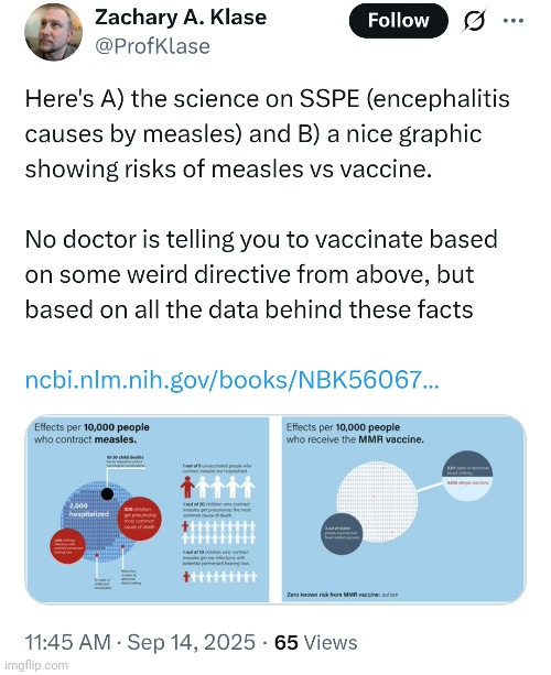
While citing a webpage may be presented as evidence, it is not the necessary scientific evidence. Therefore, simply citing a webpage does not qualify. To claim a measles “virus” causes SSPE, one must first provide scientific evidence that the measles “virus” itself exists. This requires the demonstration of purified and isolated particles obtained directly from human fluids, before any culturing or manipulation. That is the indispensable starting point for any experiment.

When pressed, Klase dodged. He reassured me that animals can be experimentally “infected” with the measles “virus” and that vaccination prevents disease in these models. But this is circular reasoning. Without purified and isolated particles as an independent variable, none of those claims hold up.


To his credit, Klase admitted he was deliberately avoiding my request, though he insisted I would just reject everything anyway.


However, that is an excuse. It is hand-waving to avoid the burden of proof. And once again, the trail went cold.
The Silence Grows Colder

So here we are, more than a month after my open challenge. Ed Rybicki has gone silent. Glenn Ison’s work seems not to exist. A retired lab tech conversation fizzled out. Zachary Klase dodged and deflected. And not one virologist has produced the absolutely necessary evidence derived from the scientific method that satisfies Koch’s Postulates proving “pathogenic viruses.” Nor has a single one presented a logical counterargument for why such evidence would be unnecessary or invalid. That should trouble anyone committed to intellectual honesty.
The longer the silence continues, the colder these trails become. And with every passing day, virologists show that they are unable to defend virology as a science, and the entire field looks more like a house of cards trembling in the frost.
2 Responses
gf7777
Mike, I would be very surprised if you hear from them. I did a peer review of TMV, but I don’t think they will accept it.
Peer Review: A Scientific Method Audit of the Tobacco Mosaic Virus Attribution Sequence
**Abstract**
This paper critically evaluates the experimental sequence that led to the institutional attribution of the Tobacco Mosaic Virus (TMV) as a causal agent of plant disease. While the TMV narrative is foundational to modern virology, a rigorous application of the scientific method reveals that key stages relied on inference, functional attribution, and model-based construction rather than structural isolation and causal demonstration. We argue that TMV was constructed from a conceptual framework, not proven to exist as a discrete, replication-competent entity. The methodology fails to satisfy core scientific requirements for falsifiability, independent variable control, and epistemic sufficiency.
**Introduction**
The discovery of TMV is widely regarded as the first demonstration of a virus. Early work by Ivanovsky and Beijerinck showed that filtered sap from diseased tobacco plants could transmit disease to healthy ones, leading to the hypothesis of an “ultrafilterable virus.” Later, Stanley crystallized a substance from infected sap that retained infectivity, and Fraenkel-Conrat demonstrated that RNA and protein components could reconstitute infectious particles. These findings are institutionally accepted as proof of viral existence. However, when evaluated against the scientific method—requiring observation, hypothesis, experimentation, falsifiability, and reproducibility—the process reveals significant epistemic gaps. This paper reviews each stage of the TMV discovery sequence and assesses whether it meets the criteria for scientific proof.
**Methodology Review**
**Stage 1: Filtration Exclusion**
Researchers filtered sap from diseased tobacco plants through porcelain to exclude bacteria and fungi. The filtrate was then applied to healthy plants, which subsequently developed symptoms. From this, it was concluded that a non-cellular infectious agent must be present. However, no structural identification of the agent was provided. Infectivity was inferred from symptom replication, not from isolation or molecular demonstration. The experiment excluded known organisms but did not positively identify a new one, violating the principle of falsifiability.
**Stage 2: Crystallization**
The filtered sap was chemically treated and centrifuged to yield a crystalline substance. This material retained infectivity when applied to healthy plants, leading researchers to declare it the virus. However, this attribution was based solely on functional outcome. There was no evidence that the crystalline substance contained a discrete, replication-competent agent. The assumption that infectivity equates to viral identity lacks structural and causal proof, and no independent variable was defined.
**Stage 3: Control Inoculation**
Healthy plants were inoculated with buffer solution alone using the same mechanical abrasion method as in the test group. Buffer-only inoculation caused minor localized damage, while application of sap from affected plants resulted in symptom patterns throughout the host. The difference in symptom severity was attributed to the presence of a presumed infectious agent in the test group. However, this attribution was based solely on symptom contrast, without structural validation of any discrete biological entity. Molecular assays, had they been performed, could only have detected specific nucleic acid sequences—not proven the existence of a replication-competent virus. No exclusion of alternative causes such as chemical irritants, RNA-induced stress responses, or mechanical trauma was conducted. The conclusion relied on functional inference—not causal demonstration.
**Stage 4: Electron Microscopy**
Purified samples from infected plants were imaged using electron microscopy, revealing uniform rod-shaped particles approximately 300 nm in length. These particles were identified as the virus. However, morphological observation alone does not establish infectivity or causal sufficiency. Without correlating these structures to molecular content and functional replication, the identification remains speculative and fails to meet the standard of causal demonstration.
**Stage 5: RNA and Protein Separation**
Researchers extracted RNA and capsid protein from material previously attributed to TMV and tested each component independently for its ability to induce symptom patterns in host plants. RNA alone was sufficient to produce symptoms, while protein alone was not. From this, RNA was identified as the genetic material responsible for symptom induction, and the capsid protein was presumed to serve a structural role. However, the experiment did not begin with a structurally isolated, replication-competent viral particle, nor did it demonstrate that the RNA and protein originated from a unified biological entity. The outcome confirmed that RNA can functionally induce symptoms, but it did not establish the existence of a discrete, causally validated virus. The conclusion rests on functional inference—not on structural isolation, replication tracking, or falsification of alternative causes.
**Stage 6: Constitution**
Researchers combined extracted RNA and capsid protein in vitro to form particles, which were then applied to healthy plants. The resulting construct produced symptoms consistent with those historically attributed to TMV. This outcome was interpreted as proof of viral existence through functional construction. However, the components used were derived from material previously assumed to contain a virus, not from a structurally isolated, replication-competent entity. No intact viral particle was demonstrated prior to assembly, and no alternative causes of symptom induction were systematically excluded. Critically, no autonomous replication cycle was observed, and no direct tracking of replication products was performed. The experiment validated a model’s ability to reproduce symptoms, but did not prove the existence of a pre-existing biological agent or demonstrate that the construct could self-propagate.
**Stage 7: Synthetic Construction**
An RNA molecule attributed to the Tobacco Mosaic Virus was chemically synthesized from a reference genome and combined with capsid protein to form synthetic particles. When applied to plants, this construct reproduced symptoms historically associated with TMV attribution. This outcome was presented as confirmation of viral existence. However, the RNA sequence was derived from a conceptual model—not from a structurally isolated, replication-competent viral entity. The reference genome itself was inferred from earlier samples that were never causally validated. No autonomous replication cycle was observed, and no direct tracking of replication products was performed. While the experiment demonstrated the model’s functional capacity to induce symptoms, it did not retroactively prove the existence of the original agent or demonstrate that the synthetic construct could self-propagate. The virus was constructed—not reconstructed.
**Replication Competence: Inference vs Demonstration**
Although an RNA molecule attributed to the Tobacco Mosaic Virus was chemically synthesized and combined with capsid protein to form a functional construct, replication competence was not directly demonstrated. The construct’s presumed ability to replicate was inferred from the appearance of symptoms in tissues beyond the inoculation site and from reproducibility of symptom patterns in other specimens following application of affected material. However, no intact viral particle was structurally validated prior to inoculation, no autonomous replication cycle was observed in vitro, and no exclusion of alternative symptom-inducing mechanisms was performed. While institutional literature describes general replication strategies for plus-sense RNA viruses like TMV—including disassembly, translation of replicase proteins, and synthesis of complementary RNA strands—these steps were not causally demonstrated in the synthetic construct. Therefore, replication competence remains a functional assumption—not a causally demonstrated property of the synthetic construct.
**Scientific Method Audit**
The TMV discovery sequence fails to meet several key criteria of the scientific method:
– Falsifiability: Early experiments did not systematically exclude alternative causes of symptoms.
– Independent Variable Control: No structurally defined, replication-competent viral particle was used as an independent variable at any stage. In synthetic construction, a model-based particle was assembled, but its structural integrity and replication competence were assumed—not demonstrated.
– Causal Demonstration: Infectivity was inferred from symptom replication, not from direct observation of replication or structural integrity.
– Reproducibility: Later stages achieved reproducibility, but only within a constructed model—not from an originally proven entity.
**Conclusion**
The TMV discovery process, though historically influential, does not satisfy the scientific method’s requirements for proving the existence of a virus. At no stage was a replication-competent viral particle structurally isolated from the original affected material. Each experimental outcome relied on functional inference, model attribution, or synthetic construction—none of which constitute causal demonstration. Replication competence was assumed based on reproducibility of symptom patterns, not directly observed or validated. The entity referred to as TMV was constructed from a conceptual framework, not proven to exist in its original biological form. This distinction is essential for preserving epistemic integrity in pathogen attribution and for ensuring that future claims of viral existence are grounded in demonstrable, falsifiable evidence.
gf7777
Mike, there is a problem that should be considered. Here’s an brief article that addresses the problem.
There’s Something Wrong With the Virologist’s Mind
Abstract
This paper examines the cognitive architecture underlying institutional virology, using the Tobacco Mosaic Virus (TMV) attribution sequence as a case study. While TMV is historically framed as the first proven virus, a forensic audit reveals that its attribution relied on functional inference, model construction, and consensus reinforcement—not structural isolation or causal demonstration. We extend this analysis to the virologist himself, treating the collective institutional mind as a diagnostic subject. We identify a systemic cognitive condition—Attribution Lock—that impairs objectivity, dissolves hypothesis discipline, and substitutes reproducibility for proof. We conclude by asking whether individuals operating under Attribution Lock pose an unintentional risk to public health, and whether safeguards should be considered to prevent harm.
Part I: The TMV Attribution Sequence—A Methodological Breakdown
The TMV narrative unfolds across seven experimental stages, each reinforcing the idea of viral existence without satisfying the scientific method’s core requirements:
– Filtration Exclusion inferred presence from symptom persistence, not structural identification.
– Crystallization equated infectivity with identity, bypassing causal proof.
– Control Inoculation used symptom contrast as exclusion logic, ignoring alternative causes.
– Electron Microscopy reified morphology without functional correlation.
– RNA and Protein Separation treated component sufficiency as entity validation.
– Constitution assembled a functional construct but failed to demonstrate replication.
– Synthetic Construction reproduced symptoms from a model-derived genome, not a proven biological agent.
Across all stages, replication competence was assumed from symptom patterns—not demonstrated through autonomous replication tracking or falsifiable experimentation.
Part II: The Institutional Virologist—A Cognitive Profile
The virologist does not approach the virus as a hypothesis to be tested. He inherits attribution as fact, and his cognition is shaped to reinforce it. His mind exhibits:
– Model Reification: Treating constructs as entities.
– Consensus Substitution: Replacing empirical proof with institutional agreement.
– Functional Inference Drift: Assuming causality from reproducible outcomes.
– Replication Assumption: Inferring biological behavior from symptom spread.
– Hypothesis Paralysis: Failing to treat foundational claims as testable propositions.
This cognitive architecture is not rare—it is institutionalized. It is trained, rewarded, and normalized across virology, diagnostics, and public health.
Part III: Attribution Lock—A Systemic Cognitive Condition
We define Attribution Lock as a cognitive condition in which practitioners treat conceptual models and reproducible outcomes as empirical proof—without structural isolation, causal demonstration, or falsifiability.
It manifests as:
– High suggestibility to institutional narratives.
– Low resistance to model reinforcement.
– Reduced epistemic skepticism.
– Reward-driven cognition aligned with consensus.
Attribution Lock is not a personal defect—it is a systemic vulnerability. But its effects are real: it disables objectivity, dissolves scientific rigor, and enables diagnostic error.
Part IV: Societal Risk—Is the Attribution-Locked Practitioner Dangerous?
While not malicious, the Attribution-Locked practitioner may pose unintentional harm:
– Diagnostic Misattribution: Treating symptoms as viral without structural proof.
– Policy Amplification: Supporting interventions based on model outputs, not causal evidence.
– Medical Overreach: Administering treatments for entities never structurally validated.
– Suppression of Dissent: Marginalizing clarity-aligned minds who demand falsifiability.
This practitioner cannot distinguish what is in his mind from what is in reality. He is epistemically hypnotized—suggestible to consensus, resistant to audit. In medical contexts, this cognitive condition may justify monitoring, restriction, or exclusion from diagnostic authority, not as punishment, but as protective oversight.
Conclusion
The TMV attribution sequence reveals not just methodological drift, but cognitive conditioning. The institutional virologist operates under Attribution Lock—a systemic impairment of objectivity and causal rigor. While not malicious, this condition may render the practitioner unfit for unsupervised diagnostic authority. We call for the development of epistemic screening tools, institutional audits, and safeguards to prevent attribution-based harm. Scientific method must be restored—not just in procedure, but in cognition.