
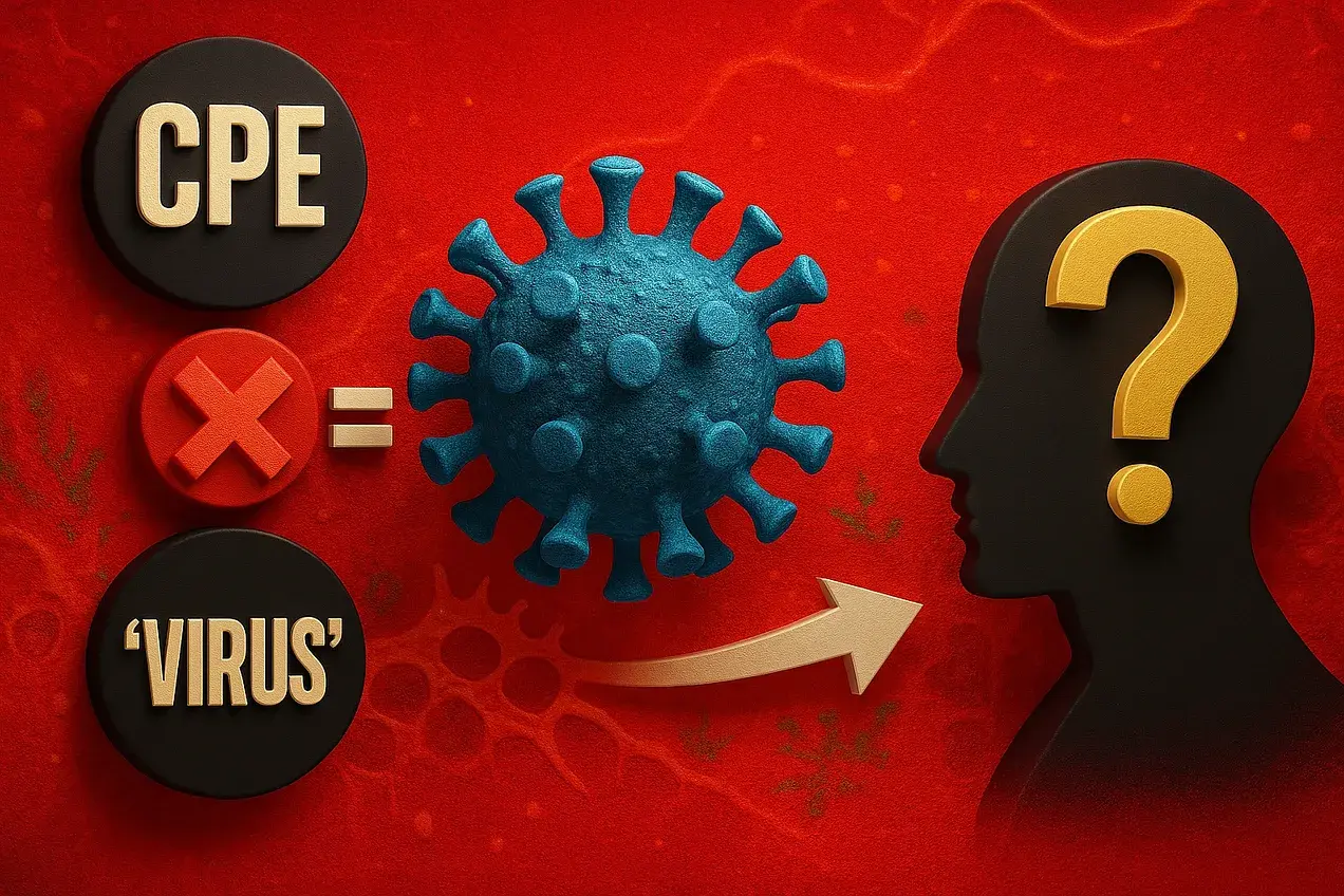
Do Cytopathic Effects Prove “Viral” Replication?
Historical Assumptions, Logical Pitfalls, and Unfalsifiable Reasoning
I want to preface this article by stating that the cell culture experiment has been repeatedly shown to be a pseudoscientific set-up. I have already written about the logical fallacies built into its design and demonstrated how it fails to adhere to the scientific method. Crucially, the experiment does not test a hypothesis derived from an observed natural phenomenon. There is no valid independent variable—no purified, isolated “viral” particles—prior to the experiment. The dependent variable, the cytopathic effect (CPE), is a non-specific, laboratory-induced artifact that can be explained by multiple causes without invoking a fictional one. Despite this, the claim that CPE equals the presence of a “replicating virus” became the cornerstone of virology. My aim here is to show that this core assumption was never proven, was baseless from the start, and has been falsified by virology’s own experiments and methods.
The advent of tissue culture techniques in the early twentieth century marked a watershed moment for the fledgling field of virology, enabling researchers to generate effects in the lab that they could then attribute to their invisible entities. Before tissue and cell culture techniques existed, “viruses” were merely a shifting concept defined purely in negative terms. As Thomas Rivers noted, they were identified by what they were not:
- invisible to ordinary microscopy,
- not recoverable through filtration,
- unable to propagate without living cells.
With the rise of tissue culture in the early 20th century, virology finally gained something visible—but only in the form of cell damage. It is crucial to note, however, that tissue and cell cultures are not natural biological systems. They are artificial laboratory constructs made from dying or weakened animal and human tissues kept alive through nutrient broths, fetal bovine serum, antibiotics, antifungals, and various chemical additives. Nothing about these mixtures resembles the internal environment of a living organism. Any effects observed within such systems reflect the breakdown, toxicity, and stress of the culture conditions themselves—not a naturally occurring process. In other words, tissue culture did not emerge from observing a phenomenon in nature and seeking its cause; instead, the phenomenon was created first, and the cause was assumed afterward.
The most striking of these lab-created phenomena were morphological changes such as rounding, detachment, syncytia formation, and eventual cell death—changes that virologist John Franklin Enders later termed cytopathic effects (CPE) in the late 1940s. These changes were quickly interpreted as proof of “viral replication.”
According to virologist Thomas Weller, who worked with Enders during the polio investigations, the so-called “viruses” induced degenerative changes in the cells in which they multiplied, changes detectable morphologically or biochemically. From the outset, these visible alterations were interpreted as direct evidence of “viral” replication, establishing a central assumption: a “virus” infects a cell, it replicates, and that replication produces CPE. This untested assumption quickly became foundational for “virus isolation,” identification, and quantification moving forward, with the Encyclopedia of Virology defining CPE as when “viruses kill or morphologically modify their host cells when they multiply.” Similarly, Fields Virology notes that “one of the classic ways of detecting virus replication in cells was the observation of changes in cell structure, or CPEs, resulting from virus infection.”
By the late 1980s, this assumption was still firmly entrenched. In the book Veterinary Virology, Frank Fenner described CPE as “visible evidence of cell damage” that occurs when “viruses” kill the cells that they replicate in:
“Many viruses kill the cells in which they replicate, so that infected cell monolayers gradually develop visible evidence of cell damage, as newly formed virions spread to involve more and more cells in the culture. These changes are known as cytopathic effects (CPE), and the responsible virus is said to be cytopathogenic.”
Similarly, Altmeyer’s Encyclopedia—a reference for medical professionals—defines CPE as the “effect of viral replication on the host cell itself.”
Taken together, these definitions show how deeply entrenched the CPE = “viral replication” assumption became. The observation of CPE was no longer treated as a phenomenon requiring explanation, but rather as an indirect marker of both the presence and multiplication of the invisible entity. Yet the critical question remained unasked: does this lab-created phenomenon actually demonstrate that “viruses” were present and replicating, or was it simply assumed to? If other evidence surfaced that refuted the CPE-equals-replication dogma, would the framework of virology even allow it to be acknowledged?
Let’s take a look.
Classical Demonstration: Enders and the “Proof” of “Viral” Replication

The work of Enders, Weller, and Robbins in the late 1940s exemplifies the historical reliance on CPE as proof of “viral replication.” According to Grafe’s A History of Experimental Virology (1994, Ch. 4), the aim of Enders’ group was to prove “viral multiplication” of the “poliovirus.” They observed that samples alleged to harbor “poliovirus” that were inoculated into human embryonal tissue produced morphological changes which were inhibited by specific “immune” serum:
“A very significant improvement that proved crucial for quantitative virology was introduced by Enders and colleagues in 1949. F.Robbins had been able to show in tissue-culture experiments in 1948 that the Lansing strain of poliovirus, which was adapted to the mouse, would multiply in a culture of human, embryonal, intestinal tissue. In an analogous procedure, virus were also successfully cultured in human embryonal nerve, skin, connective and muscle tissue. Proof of virus multiplication was obtained in subsequent infection experiments with mice and monkeys. Far more important, however, were the changes in cell morphology they could trace, and the discovery of the unstainability of infected cell nuclei. In a 1950 study, these two scientists, together with Weller, presented the “cytopathogenic effects” which were later referred to as the cytopathic effect (CPE) of cytopathogenic viruses.”
“Proof that the poliovirus, which until that time had been strictly classified as a neurotropic virus, could multiply in vitro in nonneural tissue triggered analogous tissue-culture trials with other types of virus. It should be mentioned that the aim of the Enders group was to demonstrate the multiplication of the poliovirus in the intestinal tract.”
Crucially, however, these experiments never involved purified and isolated “viral” particles. Instead, tissue homogenates alleged to contain “poliovirus” were added to cultures of human embryonal tissues, and the subsequent cellular degeneration was interpreted as “viral multiplication.” This leap—from correlation to causation—was never empirically validated.
These observations were nonetheless interpreted as confirmation that the presumed “virus” caused the observed CPE and were further used to quantify “virus” content, identify “viral” strains, and test “antiviral” substances. The trio were awarded with the Nobel Prize in 1954 based upon their polio findings, with the prize motivation being “for their discovery of the ability of poliomyelitis viruses to grow in cultures of various types of tissue.” The Nobel committee’s wording makes it clear that their interpretation of what the researchers observed (CPE in cultured cells) was framed as evidence of “viral growth/replication.” By enshrining this interpretation as a “discovery,” the award effectively erased the distinction between assumption and proof, thereby reinforcing the assumption that CPE was synonymous with “viral” growth. From that moment on, virology proceeded under a framework where visible cellular injury in culture was treated as definitive evidence of “viral replication.”
However, is this truly what Enders et al. obseeved?
Enders’ Initial Caution and Contradictions

In their 1949 paper Cultivation of the Lansing Strain of Poliomyelitis Virus in Cultures of Various Human Embryonic Tissues, Enders noted morphological differences between inoculated and uninoculated cultures after prolonged cultivation: the nuclei of cells in inoculated fragments showed a marked loss of staining properties, while cells in control cultures appeared normal. Yet, rather than leaping to a definitive conclusion, Enders wrote:
“Since the amount of material which has been studied is as yet relatively small, one cannot conclude that the changes observed in the inoculated cultures were caused by the virus.”
This admission is striking. Enders openly acknowledged that correlation did not establish causation, and that the observed cellular abnormalities might have alternative explanations. At this early stage, the link between CPE and “viral replication” was not treated as fact, but as a working hypothesis awaiting further confirmation.
But only a year later, in the 1950 paper Cytopathogenic Effect of Poliomyelitis Viruses In Vitro on Human Embryonic Tissues, Enders and his colleagues made a sweeping reversal, concluding that the cytopathic effects they observed “left no doubt” the poliomyelitis “virus” was multiplying in cells and that CPE could serve as a reliable marker for identifying “viruses.” This leap—from tentative observation to claimed certainty—became the foundation of modern virology:
‘These phenomena are of interest from two general points of view. First. they leave no doubt that poliomyelitis virus in vitro can multiply in cells other than those of the nervous system and cause profound injury of such cells. Secondly, they provide criteria by which the presence of the virus can be recognized in vitro and hence may afford a basis of technics for isolating virus from patients or animals, for the quantitative assay of virus, for serologic typing and possibly for the screening of chemotherapeutic and antibiotic substances. Further study will be required of the reliability and practicability of the application of these phenomena to such ends.”
However, only a few years later, and in the same year he received the Nobel Prize for “proving” that “viruses replicate” in tissue cultures and published his influential measles study that also showed CPE in the “uninfected” cultures, Enders published a revealing paper Cytopathology of Virus Infections: Particular Reference to Tissue Culture Studies. In it, Enders acknowledged that the central evidence—cytopathic effects (CPE)—was not specific to “viruses.” He admitted that:
- CPE can be caused by “many noxious agents,” not just “viruses.”
- Inclusion bodies are not conclusive evidence, since chemicals and unknown factors can produce them as well.
- Culture conditions and donor variables drive the outcomes, meaning age, species, and prior handling of tissue could dictate whether CPE appeared.
- Conclusions about “viral” causation were only “tentative.”
Enders wrote:
“The phenomena mentioned above under Group 1 changes may be evoked by many noxious agents. Accordingly, they cannot alone be considered as necessarily the result of viral activity. To prove this certain control procedures (serial cultivation, prevention of changes by homologous antibody, etc.) must be applied. Familiarity, however, with the effects of a specific virus in a given cell system often enables the observer to conclude tentatively that this virus is responsible.”
“Of morphological indices of viral injury, the formation of inclusion bodies (Group 2 above) is the most characteristic, although again this process cannot be accepted as conclusive evidence of viral activity since certain chemical as well as other unknown factors may condition its development. Inclusion bodies were the first cytopathic changes to be sought for in vitro and employed as criteria of infection. As indices of viral multiplication, however, they are less useful than the changes of Group 1, because these structures can be unmistakably demonstrated only in stained preparations.”
“Cytopathogenicity in vitro is influenced by factors some of which are known while many remain to be defined. At the outset a few of those now recognized will be mentioned as an introduction to the review of recorded observations on the behavior of individual agents. Of primary importance is the species from which the cells are derived. Analogous to the host range of a virus is its cytopathogenic range in cultivated cells. But correlation between susceptibility of the organism and its cells in vivo does not always exist. For although this correlation frequently obtains, the tissues of a susceptible species occasionally fail to support viral multiplication while the converse of this situation also occurs.
The age of the donor of tissue may influence cytopathogenicity. Just as young animals are frequently more susceptible to infection so their tissues may be more vulnerable to injury by the virus, yet again this correlation is not invariable. Most of the pertinent data indicate that acquired immunity to viral infections is not reflected by an increased cellular resistance, a fact advantageous from the technical point of view since it eliminates concern over the immunologic status of the donor animal.
The intensity and degree of cytopathic injury may vary according to the strain of virus or the conditions under which it has been propagated prior to its study in tissue culture. The investigator should be prepared to encounter such variations in the study of a number of representatives of a viral species. Moderate or weak cytopathogenicity may sometimes be enhanced by serial passage in vitro.”
In other words, at the exact moment virology was being elevated as having proven “viral replication,” its leading figure openly conceded that the evidence was ambiguous, confounded, and reliant on assumption.
Yet two years later in 1956, Enders framed things differently. In his Gordon Wilson Lecture, Observations on Certain Viruses Causing Exanthematous Diseases in Man, he explained that the “shortcomings” of tissue culture were overcome not by isolating and independently demonstrating a causal agent, but by observing degenerative changes in cells after exposure to suspect material:
“In this respect the shortcomings of the tissue culture have in many instances been overcome by demonstration of the fact that the majority of viruses so far examined produce, as they multiply, degenerative changes in the cells. These changes are often apparent within a few hours or a few days after the viral inoculum is introduced and frequently are sufficiently characteristic to permit a tentative identification of the virus under examination. Such effects are now referred to as “cytopathic changes” or as “cytopathogenic effects of the virus”.
Although cytopathic changes were noted earlier by various investigators, especially by Ivanovics and Hyde in 1932 and Huang in 1942, their full significance was not generally appreciated until 1950, when it was demonstrated that the viruses of poliomyelitis were highly cytopathogenic in cultures of a variety of several human tissues. Since that time results obtained with a large number of viruses and a variety of cells from various species show that exhibition of cytopathogenicity in vitro affords criteria of viral multiplication as reliable as the production of signs of infection in animals. Moreover, such criteria may be often more conveniently and accurately observed.”
Although he had previously acknowledged that CPE was nonspecific and could be due to multiple factors, Enders now elevated visual degeneration in artificial cell systems to the status of proof. He hailed the observed cell death as “criteria of viral multiplication as reliable as the production of signs of infection in animals.” These laboratory artifacts were treated as equivalent to disease itself, without first demonstrating an independently verified causal agent.
Despite having described CPE as a reliable indicator of “viral multiplication,” a year later in the 1957 paper Comments on Viral Cytopathogenicity, Enders emphasized how little was actually known about the supposed mechanism of “viral cytopathogenicity.” He openly admitted that the underlying process remained entirely mysterious and that none of the hypotheses explaining it were satisfactory—particularly because none could account for how “viral replication” could occur without cellular injury (CPE). He left it to future researchers to provide an explanation:
“In concluding these fragmentary remarks on the problem of viral cytopathogenicity, I would stress our ignorance of its underlying mechanisms. At present we simply do not know how these unique biological entities set in motion what must be an intricate chain of intracellular events that terminate in effects such as I have just described. Various hypotheses have been proposed. To me, none are entirely satisfactory, since they do not adequately allow for two of the most outstanding features I have listed, i.e., the diversity of effects produced by different viruses and the occurrence under certain conditions of viral multiplication without apparent cellular injury. But we are confident that eventually the combined efforts of biochemists and biologists will supply a full account of the factors responsible for viral disease. I venture to predict that this account will be neither simple nor soon available.”
In other words, even years after claiming to have demonstrated “viral replication,” Enders conceded that no one actually knew what caused the cellular effects being observed. The presumed “viral mechanism” remained hypothetical, and the very phenomena that earned him the Nobel Prize were still unexplained.

In retrospect, Enders himself later described in 1972 how these conclusions were formed. Reflecting on his early tissue culture experiments, he explained how they went seeking “visible evidence” that a “virus” was present in the cell culture. In essence, they assumed a “virus” was in the original sample, and then claimed that any difference between “infected” and “uninfected” cultures proved its presence:
“In 1948–1949, Doctors Weller, Robbins, and I found that the polioviruses could grow in vitro in human non-nervous tissue. This finding was particularly interesting at that time, since most people then believed that these agents were strictly neurotropic and could not multiply in other types of cells. The demonstration that this view was incorrect seemed to many to be the most significant feature of our experiments. However, we realized almost at once that although our observations might contribute to the understanding of the pathogenesis of poliomyelitis, they would have only a limited, if indeed any, practical application to diagnosis and prevention—unless we could find visible evidence for the presence of the virus within the culture itself. So, very early, we looked for changes in the cells which had been exposed to the virus. We used at first the Maitland type of culture—just fragments of chopped-up embryonic tissue. We fixed these and cut sections, examining them at different times following inoculation of the virus and comparing them with fragments from uninoculated cultures. We found suggestive indications that the virus exerted a destructive effect on the cells. We did further work on this crucial point, confirming our observations in Maitland cultures, and then went on to study the survival of such fragments and cell outgrowth after the tissue had been removed from the flask and placed in plasma drops. Finally, we exposed the monolayer outgrowth of cultures in roller tube cultures to the virus. This procedure proved to be much the best and left no doubt about the cytopathic effect which, under those circumstances, was rapid and specific, was neutralized by type-specific polio serum, and could be used to determine the infectivity titer of the virus.
This candid description reveals the circular reasoning at the heart of Enders’ approach. He and his colleagues began by presuming that a “virus” was present in the material they introduced into their cultures. Yet at that stage, they had no direct evidence that such an entity existed in the samples. Enders even conceded that the tissue-culture experiments had no practical meaning unless some visible sign appeared to confirm their assumption.
In other words, the belief preceded the evidence.
When cellular damage—cytopathic effects—eventually appeared, they interpreted it as the long-awaited confirmation that a “virus” had been present all along. But because no distinct, purified entity had been isolated beforehand to serve as the independent variable, the appearance of CPE could not logically validate their assumption. The conclusion simply restated the premise: they began by assuming a “virus,” and they ended by treating that assumption as proven.
Enders also noted that similar “cytopathic” observations had existed long before his own work but were either ignored or applied inconsistently. Early studies were frequently hampered by technical limitations, contamination, and strong interpretive bias. He openly questioned why this previously observed, laboratory-created effect had not been more thoroughly pursued, suggesting two main reasons:
- The absence of antibiotics, which made tissue culture experiments extremely vulnerable to contamination
- The dramatic nature of poliomyelitis, which finally compelled researchers to take serious notice of the phenomenon
This is a monumental confession, as it shows that the motivation for elevating artificial CPE into “viral proof” wasn’t grounded in empirical demonstration, but in a combination of practical obstacles and the emotional and sociological pressure created by a frightening disease:
I don’t know why people had for so long failed to take advantage of the fact that many viruses are cytopathogenic in vitro, because several observations of this property had been described. For example, the early ones by Ivanovic and Hyde on the virus III of Rivers; those made by Plotz and Ephrussi with fowl plague virus in the 1930s; and those reported in the early 1940s by Huang, who had made some beautiful observations on the cytopathogenicity of equine encephalitis virus in tissue cultures. Around 1939, I, with R. Relova in unpublished studies, had seen the same phenomenon—i.e., complete destruction of chick embryo cells by this agent. I think it is instructive and a bit chastening that nobody had fully exploited the potential value of the phenomenon. It was only, I believe, because of the dramatic quality of poliomyelitis as a disease that its significance became generally recognized and its potentialities widely explored. One wonders why the early observations hadn’t been followed up. Perhaps one reason was that no antibiotics were available. Thus, one couldn’t inoculate and study contaminated materials in tissue culture. Furthermore, the techniques for culture were relatively undeveloped and, as a result, the findings often were not always satisfactorily consistent. Also, I’ve always felt that the great influence of Carrel in the field was, in a sense, restrictive, because the elaborate precautions that he considered essential to avoid bacterial contamination discouraged many who otherwise might have adopted the method. But whatever the reasons may be, as we all know, it sometimes takes a long time for intrinsically important observations to become generally appreciated and applied.”
Enders’ comment about antibiotics is not a minor aside. In fact, in his 1954 Nobel Prize speech, he stated that the discovery of antibiotics “worked a revolution in the field of tissue culture.” In the Fourth R. E. Dryer Lecture Recent Observations on the Behavior in Tissue Culture of Certain Viruses Pathogenic for Man, Enders described the introduction of antibiotics as “with little doubt the most important” factor enabling the use of cytopathogenic effects as evidence of “viral” activity. Antibiotics, in effect, provided the perfect excuse—or “expectation” as Enders put it—that made it possible to claim cultures were free of everything except “viruses:”
“Today one can inoculate into tissue cultures such materials as throat washings, fecal suspensions, or preparations of organs such as intestine with the expectation that in most instances growth of larger microorganisms will not occur. Thus the presence of a virus, capable of betraying itself by manifestations such as I shall presently describe, can be recognized.”
Before this shift, investigators had no dependable internal indicator of “viral multiplication” in tissue cultures. The only way to support the presence of a “virus” was to inoculate animals with material from the cultures and observe illness or death. Even Enders acknowledged this limitation:
“Another situation that in the past limited the value of tissue cultures in the study of viruses lay in the failure of investigators to discern within the system itself a dependable indicator of viral multiplication. Accordingly, in order to demonstrate an increase in virus it was necessary to inoculate material taken from the tissue culture into a susceptible animal host. From the practical point of view, therefore, to many earlier investigators the culture seemed to offer in most instances no advantage over the animal for the cultivation of virus. Indeed, it appeared far less suitable since the culture gave no unmistakable signs of infection comparable to death or the clear emergence of pathologic changes that usually accompany the multiplication of the agents in vivo.”
Modern virology textbooks acknowledge the exact same limitation. Human Virology (4th Edition) notes that early tissue culture work was nearly impossible without antibiotics:
“But the really big breakthrough came with the discovery of antibiotics in the 1940s and 1950s. Until then, it was very difficult to keep cell and tissue cultures free from contamination with airborne bacteria and moulds, but the addition of antibiotics to the culture medium inhibited these unwanted contaminants and permitted the handling of cultures on the open bench.”
This admission is crucial. It means that the very system in which CPE became the “proof” of “viral replication” could not function without artificially modifying the environment—by suppressing bacteria, altering natural cellular behavior, and creating conditions that have no parallel in a living organism. The “evidence” for “viral replication” was inseparable from the artificial system required to generate it.
With the introduction of antibiotics, Enders and others could now assume these cultures were effectively “clean” of competing organisms. This technical change allowed the same degenerative cellular effects—once dismissed as contamination or artifact—to be reclassified as evidence of “viral” activity. The interpretive framework shifted, not because a purified causal agent had been demonstrated, but because the experimental environment had been redefined.
Structurally, Enders’ statements and contradictions reveal that the reasoning used to repurpose CPE as evidence of “viral” replication followed classic logical fallacies: affirming the consequent—assuming that because CPE occurred, “viral replication” must be occurring—and begging the question, interpreting the inhibition of CPE by “immune” serum as proof that a “virus” caused it. Without purified and isolated “viruses” serving as a true independent variable, the foundational assumption linking CPE to “viral replication” was never empirically tested.
And yet, in 1954, Enders was awarded the Nobel Prize for exactly that. The house of cards was already wobbling—just as it was being sealed into the foundations of modern virology.
Experimental Challenge: Ackermann 1954 — 1959

Interestingly, the core assumption that the observance of CPE in cultures equalled “viral replication” was challenged the exact same year Enders and his coworkers received their award for “proving” the assumption. In 1954, virologist Weston Wilbur Ackermann, who was active with polio research during the mid-20th century, published the paper Growth Characteristics of Poliomyelitis Virus in HeLa Cell Cultures: Lack of Parallelism in Cellular Injury and Virus Increase. In it, Ackermann questioned what visible cytopathology could be attributed to primary pathology or to “viral” synthesis and replication:
“Two possibilities have been considered. One is that the progressively developing visible cytopathology is the result of a progressively developing insult associated with a gradual increase in viral material. The second is that the essential injury is completed close upon the initiation of infection, in which case the visible pathology could develop independent of viral multiplication.”
Ackermann explicitly set up the hypothesis that CPE may or may not parallel “viral” replication, framing the experiments as a direct test of the classical assumption that CPE = “viral” growth. During his tests, he observed that cellular destruction occurred without any apparent “viral” increase:
Ackermann utilized methods claimed to block “viral” synthesis to further address his questions. To do so, he turned to fluoro-phenylalanine (FPA), a synthetic amino acid analog that can substitute for phenylalanine in protein synthesis. When it gets incorporated into proteins, it usually disrupts their proper folding and function. In virology research at the time, it was used theoretically as a way to inhibit “viral” replication because “viral” proteins would be malformed or nonfunctional.
Ackermann exposed HeLa cells to the FPA and found that while it acted as cytostatic (halting cell multiplication) it did not kill the cells outright. He confirmed that FPA did not directly inactivate “poliovirus,” but when cells were “infected” in its presence, the usual rise in “infectious” titer was blocked. Even so, “infected” cells still showed cytopathic changes and degenerated at the ordinary rate. In high-dose “infections,” nearly all cells succumbed as normal, but in low-dose inoculations, only a few cells developed cytopathic effects and the damage did not spread to neighboring cells. Normally the “infection” would propagate through the culture, but here it remained confined, which was taken as evidence that new “virus” was not being produced and transmitted.
From this, Ackermann drew the conclusion that “viral replication” and cellular damage were separable phenomena—i.e., the CPE was not strictly dependent on “virus multiplication.” He described the processes as having “a significant degree of autonomy.” In other words, the experiment suggested that whatever causes the cell damage (what virologists interpreted as “viral cytopathogenicity”) was not simply the same as “viral replication.” That result undercut the assumption that cell death in culture = “virus replication” = proof of “viral activity:”
“While the fluoro derivative completely inhibited viral multiplication, it did not prevent the cytopathogenic effect of the virus. In the presence of fluoro-phenylalanine, the disintegration of an infected cell proceeded at what appeared to be the ordinary rate, without any increase of the infectious agent. Experimentally the processes leading to viral increase and to cellular injury have been shown to possess a significant degree of autonomy.”
This is a direct experimental falsification of the assumption that CPE indicates “viral replication” using their own standards and methodology.
Ackermann also described cytoplasmic granules and nuclear changes as a “striking” and consistent feature of “poliovirus infection” in HeLa cells, claiming they weren’t present in controls. Yet in the same breath he admitted they sometimes appeared after chemical injury or exposure to “toxic” stool suspensions. That admission destroys the claim of specificity: if the same morphological effects can arise from “non-viral” insults, then they cannot serve as proof of a unique “viral” cause. At best, they show a nonspecific cellular stress response misinterpreted as evidence of “viral” activity:
“A culture 21 hours after infection is shown in Fig. 4. Most of the cells no longer adhere to the coverslip. The few that do show marked nuclear pyknosis, and a striking finding is the presence of many deep blue-staining cytoplasmic granules which are irregular in size and shape. These are consistently found in HeLa cells undergoing cytopathology after infection with a number of strains of Type I and Type III poliomyelitis virus. They have not been seen in control cultures, and only rarely in cultures incubated for long periods without change of medium or in cultures injured by various chemical agents or “toxic” stool suspensions.”
Ackermann’s findings show that cell death in culture can arise independently of “viral replication” and can be mimicked by toxins or chemical injury. Far from confirming a “viral” cause, CPE is better understood as a nonspecific stress response that is misinterpreted as proof of “viral” activity.
In a follow-up study from 1958 titled Concerning the Cytopathogenic Effect of Poliovirus; Evidence for an Extraviral Toxin, Ackermann confirmed these findings, showing that cellular injury and “virus” production were autonomous processes, directly falsifying the assumption that CPE proves “viral replication.” Unfortunately, the paper is behind a paywall, but some perspective about the work can be gained from the available abstract:
“A toxic effect of fluids obtained from tissue cultures infected with type 1 poliovirus was observed when the fluids were introduced into fresh cultures. The destructive activity is distinct from the usual virus-induced cytopathogenic effect in that it occurs rapidly, without a prolonged latent period and without virus production. The infectious activity can be selectively neutralized by antiserum with full retention of the toxicity. However, certain antisera will also neutralize the toxin. Further, the two activities possess differential heat stability. The ability to destroy cells and give rise to detectable amounts of toxin is a characteristic of only certain lines of the poliovirus.”
Fortunately, a few more details on Ackermann’s work can be found at the Cabi Digital Library. The researchers described how the loss of cells in “poliovirus-infected” cultures occurred in two distinct phases: an early wave of cell destruction within 2–4 hours, before any detectable “virus” appeared inside cells, and a later wave at 6–12 hours, coinciding with the supposed appearance of “intracellular virus.” The early destruction was attributed to a toxin-like factor present in the culture fluid. Crucially, the toxic activity behaved differently from “infectivity:” while “virus infectivity” remained stable for weeks at 4°C, the toxicity decayed rapidly, showing they were separable phenomena. Antisera experiments reinforced this distinction—some sera neutralized “infectivity” but not toxicity, while others blocked both until adsorbed with HeLa cells, after which only “infectivity” was inhibited.
These results show that CPE was not an inevitable consequence of “virus multiplication” but instead could stem from unstable, cell-derived toxic byproducts. Far from proving “viral replication,” Ackermann’s findings exposed the ambiguity of culture-based assays and suggested that what virologists labeled “cytopathogenicity” was often a nonspecific toxic effect of their experimental system:
“A toxic action separate from infectivity has been described for a wide variety of viruses. This paper describes experiments suggesting that tissue cultures infected with poliovirus contain a toxin as well as infectious virus:
1. The loss of cells from a culture infected with a large dose of poliovirus (Type 1 Mahoney PT strain) occurs in 2 stages: – (a) at 2-4 hours after infection, before the appearance of intracellular virus; (b) at 6-12 hours, after the appearance of intracellular virus. The first of these periods is thought to be associated with viral toxin.
2. There is little or no loss of infectivity when poliovirus is stored for several weeks at 4°C. but the toxic activity rapidly declines.
3. Antisera can be obtained which will only neutralize virus infectivity, not toxicity. Thus a monkey antiserum to Brunhilde Type 1 virus neutralized virus, but not toxicity. Similarly an antiserum made in monkeys with fluid from HeLa cell cultures infected with Type 1 inhibited both infectivity and toxicity, but after adsorption with HeLa cells only affected infectivity. The action against toxicity was probably not due to antibodies directed against HeLa cells since.
(a) another antiserum against HeLa cells was not effective,
(b) HeLa cell extracts were not toxic.
4. Not all strains of poliovirus appear to produce toxin. Thus the PT strain (a large-plaque former passed in HeLa cells) of Mahoney Type 1 poliovirus produced toxin in HeLa and monkey kidney cells whereas the pt strain (a small-plaque former passed in monkeys and in monkey kidney cells) also of Mahoney Type 1 did not produce toxin in either type of cell culture.”
Ackermann’s results are exactly what you’d expect if CPE were driven by unstable, toxic byproducts of stressed or dying cells, not a replicating microbe. The instability of the toxin compared to “infectivity,” its variability across strains and passages, and its selective neutralization by antisera all reinforce that this was a nonspecific cellular toxicity. Instead of reinforcing CPE as an indicator of “viral replication,” Ackermann’s work exposed the fatal flaw in virology’s foundation: CPE is not evidence of a “virus.”
In the 1959 paper Biochemical studies of virus-infected cells, Ackermann further undermined the “viral-replication” narrative by pointing out that most of the new material produced in so-called “virus-infected” cells was not “viral” at all. Based on its sheer quantity, subcellular distribution, and RNA composition, he noted that the bulk of the products formed during “poliovirus infection” were clearly cellular in nature, with similar findings reported for “herpesvirus” and “adenovirus.” In fact, biochemical studies revealed elevated levels of several glycolytic enzymes—normal metabolic proteins—appearing in the same cytoplasmic fraction where increased protein content had been measured. These observations suggested once again that what virologists were interpreting as “viral synthesis” was more likely a nonspecific cellular stress response or metabolic derangement, not the creation of new “viruses.”
“It is quite clear from experimental evidence (the quantities of materials involved, their distribution among the subcellular components, and the base composition of the RNA) that much material formed during poliovirus infection is indeed not of the viral type (Maassab et al., 1957; Loh et al., 1958). On the basis of quantitative considerations alone, it would appear that similar results obtain from infection with herpes virus (Newton and Stoker, 1958) and with adenovirus (Boyer et al., 1957). The second point, regarding the cellular nature of the newly formed material, awaits further investigation. However, in the case for poliovirus, the recent report of elevated levels of six glycolytic enzymes following infection (Matzelt et al., 1958) is of particular interest when we consider that normally they are found in the soluble fraction of the cytoplasm. This corresponds to fraction 111, which we have found to increase in total protein content (Loh et al., 1958).”
Ackermann admitted CPE may not result from a “virus” at all, but from the cell’s own unbalanced growth or toxic byproducts accumulating in culture. In other words, the very marker virologists rely on is non-specific and indistinguishable from chemical injury:
“From this line of reasoning, the difficulty arises when the normal activity stimulated by the virus is out of phase with other normal activities of the cell. The cytopathogenic effect might have its primary basis, not in the synthesis of a small amount of foreign material, but in an unbalanced growth of the cell. However, the final loss of morphologic integrity may result from the secondary accumulation of some particular substance. Such a cytotoxic substance has been described in a preliminary publication (Ackermann et al., 1958~).”
Taken all together, Ackermann’s work showed, using virology’s own methods and standards, that CPE is not a reliable indicator of “viral replication.” Instead, it can just as easily be explained by disrupted cellular processes and toxic byproducts of the culture system itself. His critical findings—particularly the identification of a toxin-induced cytopathic effect—had a lasting influence on how virologists interpret culture-based assays and the biochemical underpinnings of “viral infection.” Crucially, they exposed how nonspecific and misleading the culture assay really was.
Emergence of Unfalsifiable Reasoning

Following these experimental challenges, it became clear that CPE could occur in the absence of demonstrable “viral replication,” and, as Enders himself acknowledged, that “viral replication” could occur without observable CPE. Therefore, virology developed a series of interpretive “rescue devices” to preserve the prevailing hypothesis. Whenever cytopathic effects (CPE) failed to align with expectations, new categories such as “non-cytocidal infection,” “persistent infection,” “abortive infection,” or “host-cell variability” were invoked. As virologist H.G. Pereira admitted in The Cytopathic Effect of Animal Viruses, even the core link between CPE and “viral replication” was inconsistent:
“Cytopathic action is a common manifestation of virus infectivity and is usually accompanied by synthesis of fully infectious virus particles or of noninfectious virus materials. However, CPE occurring in the absence of detectable production of virus materials and, conversely, virus multiplication in the absence of evident CPE, have been observed in a number of systems.”
This acknowledgment was striking: CPE could occur without any “virus” being detected, and “virus multiplication” could allegedly occur without any visible cytopathic effect. In either case, the causal inference was preserved through reinterpretation rather than empirical confirmation.
Even more telling, Pereira documented that so-called “viral CPE” was not a fixed, autonomous signature, but could be induced or suppressed simply by altering culture conditions. For example, Bang et al. (1957) reported that recovery from supposed “infection” with Eastern equine encephalomyelitis “virus” was favored when embryo extract was added to the medium and incubation occurred at 37°C, whereas maintenance of chronic “infection” required both a lower temperature and the same embryo extract. Likewise, Ginsberg (1958) and Takemoto & Habel (1959) showed that use of deficient media led to increased CPE, which was then equated with greater “virus production.” Pereira further noted that primary tissue cultures displayed a “gradual increase in susceptibility to viral CPE” simply as they aged (Kumazai et al., 1958; Frothingham, 1959; Chaproniere, 1960). In other words, the presence, absence, or severity of CPE could be toggled by nutrient availability, incubation temperature, additives, or the age of the culture—factors entirely external to any alleged “virus.”
Pereira ultimately conceded in his conclusion that no CPE is uniquely diagnostic of any so-called “virus:”
“An attempt to characterize the CPE of major virus groups is presented in Table I. It must be stressed that none of the characters listed in this table is pathognomonic of infection by any given virus, and that exceptional behavior may be shown by individual viruses under certain conditions. However, provided these characters are considered as parts of the over-all picture of cytopathic action, they furnish valuable evidence suggesting or supporting the existence of possible relationships between different viruses.”
This is an extraordinary admission. Not only is no cytopathic effect pathognomonic (i.e., sign that by itself establishes the diagnosis), but exceptions always appear, and the results can only ever “suggest” relationships rather than prove causation. In other words, the very phenomenon once treated as definitive evidence of “viral replication” was admitted to be nonspecific, inconsistent, and open to reinterpretation.
Modern virology textbooks echo these same concessions. Human Virology (4th Edition) openly describes “viruses” that produce no visible CPE, are detected only by indirect methods such as “superinfection resistance” or immunofluorescence, and in some cases cannot be grown in cell culture systems at all.
“A few viruses, although replicating in the cell culture, cause no visible CPE and are detected only by their ability to make the cells resistant to superinfection with a second virus. Other viruses not causing CPE can be detected by immunofluores- cence (Fig. 36.1) or by their capacity to bind red blood cells (haemadsorption). Some viruses, e.g. certain enteric and hepatitis viruses cannot be grown in cell culture systems.”
The authors even note that “isolating HIV” required artificially stimulating lymphocytes with plant lectins and IL-2 because lymphocytes “cannot normally be maintained in culture.” In other words, even according to mainstream virology, the phenomenon once treated as the primary evidence for “viral replication” was never universal, reliable, or self-validating.
The American Society for Microbiology admitted, in their document Cytopathic Effects of Viruses Protocols, that many “viruses” cause no CPE at all in their alleged natural host cells:
“Some viruses cause very little or no CPE in cells of their natural host. Their presence can be detected visually only by hemadsorption or interference, in which infected cell cultures showing no CPE inhibit the replication of another virus subsequently introduced into the cultures, or in situ by viral antigen or nucleic acid detection.”
They also caution that there is no fixed standard for what CPE should look like:
“Keep in mind that a given virus may not conform to the norm for its family, or it may produce different CPE in different host cell types.”
Indirect detection methods such as hemadsorption, interference assays, and molecular techniques were employed when CPE was absent. Principles of Molecular Virology by Cann explicitly acknowledges the historical role of CPE as a proxy for replication and the eventual reliance on genetic analysis as a stand-in when it does not occur:
“In the case of viruses for which no such systems exist—because they are not cytopathic, or do not replicate in culture, or did not cause local lesions—little genetic analysis was possible before the development of molecular genetics, whereafter progress was extremely rapid in many cases.”
Likewise, Fields Virology conceded the same point—other methods must be utilized when there is a failure to observe CPE:
“Cytopathic effect is the simplest and most widely used criterion for infection, but not all viruses cause a cytopathic effect, and in these cases other methods must suffice.”
It further admitted that “viruses” can grow to high titer without producing CPE, necessiting other means of detection:
“Viral growth in cell cultures is most often detected based on the development of microscopically visible cytopathic effect (CPE). Some viruses, however, can grow to high titer without producing visible CPE and must be detected by other means.”
This was not just a theoretical concession; it shaped day-to-day lab practice. For example, Minamoto et al. (1976) describe using hemadsorption as a surrogate indicator for rabies “virus” growth:
“The HAD phenomenon was applied to rabies SN test as indicator of virus growth instead of cytopathic effect (CPE).” (Journal of Virology, 20(1): 234–241).
In other words, the absence of cytopathic destruction never falsified the hypothesis. Instead, the evidentiary net was expanded: any observable change could be reinterpreted as “viral replication,” and the lack of CPE was explained away by invoking alternative mechanisms or indirect molecular surrogates. Over time, the center of gravity shifted from direct, visible effects (already ambiguous and artificial) to biochemical traces and genetic fragments—which themselves presuppose the very entities they were supposed to prove.
These strategies rendered the hypothesis unfalsifiable: any CPE pattern could be retroactively rationalized as proof of “replication,” and any absence of CPE could be explained away by appealing to alternative mechanisms or indirect surrogates. By this logic, no possible outcome could ever count as evidence against the existence or activity of a “virus.” The result was that virology effectively insulated its core assumption from empirical falsification.
Logical Pitfalls and Unfalsifiable Reinforcement

The trajectory of CPE reasoning illustrates how classical virology entrenched itself in a web of logical pitfalls and interpretive escape hatches. Although cytopathic effects had been observed before Enders, these findings were largely dismissed as contamination, technical failure, or laboratory artifact. What changed was not the phenomenon itself, but the interpretive environment. With the advent of antibiotics—and under the intense social and political urgency to combat poliomyelitis—Enders was able to reframe this previously disregarded laboratory effect as meaningful biological evidence for the presence of a “virus.” A phenomenon once treated as noise was repurposed as proof.
From that point forward, cytopathic effect (CPE) was treated as evidence of “viral replication,” despite the fact that this inference was never demonstrated using purified “viral” preparations. The reasoning relied on classic logical fallacies. Because “viruses” were presumed to cause cell damage, the observation of cell damage was taken as confirmation that a “virus” must be replicating—an instance of affirming the consequent.
This circularity was reinforced through the use of “immune” serum: when “antibodies” inhibited CPE, this was treated as proof that a “virus” had caused it, even though the causal chain had never been independently established. When CPE failed to appear, the underlying assumption remained intact, and alternative “viral” explanations were introduced to preserve it. In all cases, the existence and activity of the “virus” were assumed rather than demonstrated.
As contradictions grew, the goalposts shifted. CPE was initially treated as the defining criterion, but when exceptions mounted, the definition of “viral replication” was broadened to include hemadsorption, interference, antigen staining, or genetic fragments. No matter the outcome, “replication” was always said to be occurring.
The result was a framework designed to resist falsification. For a hypothesis to be scientific, some observation must exist that could prove it wrong. Yet here, both the presence and absence of CPE, as well as any indirect biochemical marker, were all reinterpreted as evidence of replication. By design, the assumption could not be refuted, only reinforced.
The history of CPE highlights a larger lesson about how science can go astray when assumptions are protected rather than tested. This unfalsifiable structure was reinforced by authoritative awards and textbooks, ensuring that generations of researchers inherited the assumption rather than question it. The case of CPE shows how easily correlation can be mistaken for causation, and how logical fallacies can become institutionalized as scientific method. Historical reliance on CPE, bolstered by post hoc rescue devices, entrenched an unfalsifiable doctrine supported by unsound reasoning. This underscores the necessity of independent verification and genuine falsifiability in science, showing how an untested assumption can harden into dogma and shape decades of practice.
Virology failed to confront its flawed foundational reasoning and bypassed the principle that true science demands: hypotheses must be testable, and they must be vulnerable to being proven wrong. From its earliest days, the field substituted assumption for demonstration. Effects were treated as causes. Absences were repackaged as presences. And circular logic was elevated to methodological standard.
In that light, the repurposing of cytopathic effects as stand-ins for “viral” existence and replication is not a triumph of science but a symptom of its absence. Virology did not drift away from empirical rigor—it was built without it.
3 Responses
J.P
For some reason your coloured quote boxes transform into the most obnoxious navy blue and deep purple in my email inbox that are truly horrendous to read. I can’t explain the discrepancy between the paler, easier-on-the-eye colours here and what comes through via email.
Great article, though!
Mike Stone
Hi J.P.,
That’s frustrating to hear about the color change. Unfortunately, that would be on the WordPress side. I don’t believe I have any control over it. Thanks for bringing it to my attention.
gf7777
Thanks Mike. This information always comes in handy, the detail is especially valuable.
Bad news:
USDA Fast-Tracks ‘Experimental’ Self-Amplifying m-RNA Injections for Cats and Dogs Without Safety Testing
https://thepeoplesvoice.tv/usda-fast-tracks-experimental-self-amplifying-mrna-injections-for-cats-and-dogs-without-safety-testing/
I made a brief article about this and posted it on numerous subreddits. On two of them it was removed fairly quickly. It’s really a sad thing about these moderators don’t want people to understand the basic facts. And that’s all this article presents. But they don’t want to hear it. I still feel sorry for their pets. People are stupid.
New Vaccines for Pets
What’s Being Injected and What’s Assumed
A new kind of injection is now being used in veterinary medicine. These products are called mRNA vaccines. They are being introduced for conditions such as canine flu, feline leukemia, and rabies. These are not traditional vaccines. They are based on a different theoretical process—one that relies not on direct observation of biological systems, but on models of how those systems are believed to function.
Traditional vaccines are assumed to work by exposing the body to weakened or inactivated forms of what virology identifies as a virus. The immune system is expected to respond to this material and develop a kind of memory. mRNA vaccines do not follow this approach. Instead, they contain synthetic genetic material—messenger RNA—encased in lipid nanoparticles. These particles are injected into the body with the intention of entering cells and delivering the mRNA. According to the model, the cell reads the mRNA and produces a specific protein. That protein is then expected to trigger an immune response.
This process is not directly observed. It is inferred from a model of cellular function. That model is built from laboratory procedures such as cryo-electron microscopy, X-ray crystallography, and other techniques that require freezing, slicing, staining, or chemically altering biological material. These methods do not show the living cell in its original state. They produce images and data that are interpreted through assumptions about what the cell is and how it works. The structures and processes described in textbooks are not photographs of reality. They are reconstructions.
The mRNA in the vial is real. The lipid nanoparticles are real. But what happens after injection is not directly known. It is projected from a model. If that model is incorrect or incomplete, the outcome may not match expectations. The synthetic mRNA may interact with cells in ways that were not predicted. The lipid nanoparticles may distribute throughout the body and enter tissues that were not intended targets. The immune system may respond in ways that are not beneficial. These are not theoretical risks. They are physical possibilities that follow from the fact that the intervention is real, while the system it acts upon is not fully understood.
When scientists refer to the mRNA producing a “viral protein,” that language also comes from the model. The concept of a virus, and the specific proteins it is said to contain, are not directly observed entities. They are inferred from patterns in cell cultures, genetic sequences, and laboratory effects. Virology and biology rely on each other’s models to support their claims. Biology provides the model of the cell. Virology defines the virus in terms of how it is thought to interact with that cell. These models reinforce each other, but they do not confirm each other. They do not provide direct evidence of the original condition of the material being studied.
This is especially important in veterinary medicine. Animals cannot describe how they feel. They cannot report side effects. Long-term studies are limited. Many pet owners may not be aware that the injection being offered is based on a new and largely untested technology. The assumption may be that it is just another routine shot, when in fact it represents a major shift in how biological intervention is being approached.
The use of mRNA injections in animals is not simply a scientific development. It is a material application of a theoretical framework. That framework may or may not reflect the true nature of the living systems it claims to describe. The distinction between what is real and what is modeled remains unresolved.