
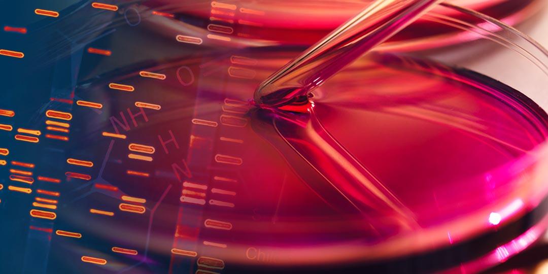
The Cell Cuture Problem
“Cell culture relies on the assumption that the behavior of cells in vitro is fundamentally similar to their behavior as part of a tissue within an organ of a multicellular organism.”
Is it time to reinvent basic cell culture medium? (physiology.org)
When virologists claim they have isolated a “virus,” they do not mean that they separated a particular particle they believe to be one from everything else. What they mean is that they took some fluid from a sick patient, added it to a cell culture typically consisting of african green monkey kidney (VERO) cells, antibiotics/Antifungals, fetal bovine serum, DMEM “nutrients,” etc., and then left the toxic mixture to incubate for days/weeks/months to see if they observe what they call cytopathic effects, which is nothing more than cellular breakdown assumed to be caused by a “virus.”
For example, this is the detailed method of “virus isolation” from one of the original “SARS-COV-2” papers:
A pneumonia outbreak associated with a new coronavirus of probable bat origin
Sample collection
“Human samples, including oral swabs, anal swabs, blood and BALF samples were collected by Jinyintan hospital (Wuhan, China) with the consent of all patients and approved by the ethics committee of the designated hospital for emerging infectious diseases. Patients were sampled without gender or age preference unless indicated. For swabs, 1.5 ml DMEM containing 2% FBS was added to each tube. The supernatant was collected after centrifugation at 2,500 rpm, vortexing for 60 s and a standing period of 15–30 min. The supernatant from swabs or BALF (no pre-treatment) was added to either lysis buffer for RNA extraction or to viral transport medium for isolation of the virus. The viral transport medium was composed of Hank’s balanced salt solution (pH 7.4) containing BSA (1%), amphotericin (15 μg ml−1), penicillin G (100 units ml−1) and streptomycin (50 μg ml−1). Serum was separated by centrifugation at 3,000g for 15 min within 24 h of collection, followed by inactivation at 56 °C for 1 h, and was then stored at 4 °C until use.
Virus isolation, cell infection, electron microscopy and neutralization assay
The following cell lines were used for virus isolation in this study: Vero E6 and Huh7 cells, which were cultured in DMEM containing 10% FBS. All cell lines were tested and free of mycoplasma contamination, submitted for species identification and authenticated by morphological evaluation by microscopy. None of the cell lines was on the list of commonly misidentified cell lines (by ICLAC).
Cultured cell monolayers were maintained in their respective medium. The PCR-positive BALF sample from ICU-06 patient was spun at 8,000g for 15 min, filtered and diluted 1:2 with DMEM supplemented with 16 μg ml−1 trypsin before it was added to the cells. After incubation at 37 °C for 1 h, the inoculum was removed and replaced with fresh culture medium containing antibiotics (see below) and 16 μg ml−1 trypsin. The cells were incubated at 37 °C and observed daily for cytopathogenic effects. The culture supernatant was examined for the presence of virus by qRT–PCR methods developed in this study, and cells were examined by immunofluorescence microscopy using the anti-SARSr-CoV Rp3 N antibody that was generated in-house (1:1,000). Penicillin (100 units ml−1) and streptomycin (15 μg ml−1) were included in all tissue culture media.”
https://www.nature.com/articles/s41586-020-2012-7
As you can see, these cultures are a mixture of MANY different chemicals and nutrients and to believe that these have no effect on the cells causing the CPE claimed to be due to a “virus” is absurd. We know for a fact that these chemicals do alter the cell and potentially anything contained within the culture:
Antibiotics:

Antibiotics in Cell Culture: Friend or Enemy?
“This is why standard cell culture protocols often include the prophylactic use of antibiotics, such as penicillin, streptomycin, gentamicin or amphotericin as media supplements to reduce infection rates. Relatively little is known, however, about the effects of these substances on the metabolism of cultured cells, cell proliferation, differentiation or gene expression. Do antibiotics indeed help to solve the contamination issue, or are they creating additional new problems? “The main goal to use antibiotics in cell culture is to kill bacteria or inhibit their proliferation,” explains Dr. Muna Ali, a scientific support specialist at PromoCell. “However, it is easy to forget that they can also harm the cells themselves as they cause many side effects: For instance, antibiotics can attack other specific, non-bacterial structures in the cell.”
“Llobet and colleagues described how the mix of penicillin/streptomycin or gentamicin alone can affect the differentiation of human adipose-tissue derived stem cells into adipocytes. Similar effects were observed on embryonic stem cells (Cohen et al., 2006, Varghese et al., 2017), mesenchymal stem cells (Chang et al., 2006), primary cancer cell lines (Relier et al., 2016) and keratinocytes (Nygaard et al., 2015). The use of antibiotics can also significantly alter gene expression and regulation (Ryu et al., 2017) and could modify the results of studies focused on drug response, cell cycle regulation and cell differentiation.”
“The parallel test showed conclusively that cells treated with antibiotics grow slower and stop proliferating earlier,” says Ali.”
“When culturing NHEK, we add penicillin and streptomycin to avoid microbiological contamination. However, we realized that the cells showed a poor growth rate and adherence to the culture dish in the presence of antibiotics.” Parallel tests like the ones in Haarmann-Stemmann’s lab also confirmed these findings: The adverse effects of antibiotics might be caused by their negative influence on mitochondrial functions. According to the endosymbiont theory, mitochondria are of bacterial origin, and their molecular and structural components are very similar (Singh et al., 2013).
In sum: Try to exclude antibiotics from your cell culture
“The above-mentioned findings strongly confirm PromoCell’s recommendation of avoiding the use of antibiotics in cell culture,” concludes Ali. “Most primary or normal human cells show reduced growth rates in the presence of antibiotics. Keeping the cells free from microorganism contamination can be accomplished with proper knowledge of good laboratory practice. Following all the guidelines towards a sterile technique makes these compounds unnecessary.”
https://www.promocell.com/…/antibiotics-in-cell…/
Fetal Bovine Serum:

Fetal Bovine Serum RNA Interferes with the Cell Culture derived Extracellular RNA
“Fetal bovine serum (FBS) has been used in eukaryotic cell cultures for decades. However, little attention has been paid to the biological effects associated with RNA content of FBS on cell cultures. Here, using RNA sequencing, we demonstrate that FBS contains a diverse repertoire of protein-coding and regulatory RNA species, including mRNA, miRNA, rRNA and snoRNA. The majority of them (>70%) are retained even after extended ultracentrifugation in the preparations of vesicle-depleted FBS (vdFBS) commonly utilized in the studies of extracellular vesicles (EV) and intercellular communication. FBS-associated RNA is co-isolated with cell-culture derived extracellular RNA (exRNA) and interferes with the downstream RNA analysis. Many evolutionally conserved FBS-derived RNA species can be falsely annotated as human or mouse transcripts. Notably, specific miRNAs abundant in FBS, such as miR-122, miR-451a and miR-1246, have been previously reported as enriched in cell-culture derived EVs, possibly due to the confounding effect of the FBS. Analysis of publically available exRNA datasets supports the notion of FBS contamination. Furthermore, FBS transcripts can be taken up by cultured cells and affect the results of highly sensitive gene expression profiling technologies. Therefore, precautions for experimental design are warranted to minimize the interference and misinterpretations caused by FBS-derived RNA.”
https://www.nature.com/articles/srep31175
DMEM (and Other Media):

Is it time to reinvent basic cell culture medium?
“Notably, none of the examined media fully adhere to physiological values of electrolytes and carbohydrates (Table 1). The most commonly used media, DMEM and RPMI 1640, deviate the most from physiological values. DMEM contains 25 mM glucose, four times more than normal and rare even in hyperglycemia (26). Sigma carries a low-glucose variant of DMEM; however, a Google Scholar search suggests that it was utilized in only 2,400 of 31,300 studies using DMEM in 2016. RPMI 1640 contains extremely low levels of calcium, magnesium, and sulfate, elevated glucose at 11 mM, and threefold higher than normal phosphate levels. Compared with the physiological levels in human plasma, DMEM and MEM have higher calcium, MEM and M199 have higher chloride and sulfate, and M199 also has slightly increased sodium and potassium.”
“The important question is whether the observed deviations in electrolytes and glucose can impact cell behavior.”
“In conclusion, we suggest that the nonphysiological electrolyte and carbohydrate microenvironment of the most commonly used cell culture media may result in unintended and uncontrolled changes in cell behavior, thereby contributing to the difficulties in reproducibility observed in modern publications (4). In contrast to finding technological solutions for imitating mechanical environment, oxygen levels, and minimal biological supplements, changing ion and glucose concentrations to better model biological fluids is easy, and only requires a collective decision of scientists and industry. It is time to make such a decision.”
https://journals.physiology.org/…/ajpcell.00336.2016
How can virologists claim that what is occuring in cell cultures is natural and not affected by all the added ingredients/chemicals/nutrients? It is absurd to believe the original sample and the cell are unaltered. These cell cultures are NOT PURIFIED unaltered particles that are separated from everything else. They are toxic concoctions for which a “virus” is ASSUMED to exist within.
0 Responses
There are no comments yet. Be the first to leave a response.