
“There is a debate tactic known as ‘elephant hurling’. This occurs when the critic throws summary arguments about complex issues to give the impression of weighty evidence, but with an unstated presumption that a large complex of underlying ideas is true, and failing to consider opposing data, usually because they have uncritically accepted the arguments from their own side. We should challenge elephant-hurlers to offer specifics and challenge the underlying assumptions.”
Excerpt from Refuting Evolution 2 by Jonathan Sarfati, Ph.D. with Michael Matthews
To those who have looked into virology both critically and logically, it is painfully obvious that there is no scientific evidence for the existence of any so-called “virus.” It simply does not exist. This holds equally true for the most recent fictitious boogeyman in “SARS-COV-2.” No “virus” has ever been properly purified nor isolated directly from the fluids taken from a sick host and then shown to be pathogenic in a natural way while adhering to the scientific method in order to demonstrate cause and effect. As no “virus” has ever been scientifically proven to exist, it should go without saying that no spike protein said to belong to one has ever met this same criteria either. I recently went through this lack of evidence for the “coronavirus” spike protein here. Thus, it is honestly a waste of time to even entertain the idea of a spike protein until such evidence can be produced for both the “virus” in question and its assumed proteins.
So why am I devoting another article to this 9-12 nm figment of the imagination? I was recently sent a post by Jeremy Hammond titled: Fact Check: COVID-19 Vaccine mRNA and Spike Protein Are Not Cleared ‘Within Days’ which was supposed to be fact-checking the “fact-checkers.” In this article, Mr. Hammond attempted to debunk a “fact-check” by Health Feedback which itself aimed to allay fears about the spike protein causing damage to the body after vaccination. According to “Covid” mythology, the spike protein is claimed to circulate inside the body after mRNA injection. After submitting oneself to the toxic experiment, an unobservable magical process is said to occur where the invisible mRNA puts on its teaching cap and instructs the body on how to create the invisible spike proteins in order to learn how to develop an immune response so that it can fight off the spike protein that it just discovered how to create. Seems like a “beLIEvable” story about the novel “virus,” right?
The Health Feedback “fact-check” argued that the spike protein is perfectly safe as it is produced in such small quantities in the body that it is harmless. Mr. Hammond, on the other hand, disagreed with their story and proceeded to provide his own version of events in order to claim that the spike protein by itself is harmful. While it is not my intent to make the case that the Health Feedback “fact-check” is correct (it most definitely is not) nor that the mRNA injections are safe by any means (I have previously shown that they are in fact very harmful with many unknown side effects), I must interject here as Mr. Hammond is attempting to debunk one fictional narrative with yet another, both of which are tied to the existence and supposed pathogenicity of a spike protein.
From what I’ve been told, Jeremy Hammond is for health freedom and he has a large following. Thus, it is very important that he gets his facts straight so as to not mislead people down a side road right back into the pharmaceutically-endorsed germ theory lie. While I do not know much about him, I have been told that, while he has consistently and rightfully called out the dangers of the vaccines, Mr. Hammond very much pushes the myth that “viruses” exist. That much is clear from his article insinuating that such a thing as a spike protein exists and is by itself pathogenic. Now, it is not my intent to attack Mr. Hammond. Rather, this is an attempt to set the record straight based on the evidence available. Hopefully, this will also stand as a good lesson in always vetting any study before submitting it as evidence to bolster your own argument.

While reading the article, it became clear that Mr. Hammond was using an unsavory tactic to try and debunk the Health Feedback “fact-check.” This tactic is known as elephant hurling, which is where one throws out numerous arguments and/or studies in order to create the illusion that the weight of the evidence is on their side. It is an intimidation tactic designed to overwhelm not only the “opponent” (Health Feedback in this case if it were an actual debate) but also those reading the article. It is an attempt to claim that a preponderance of evidence means that one’s argument is correct. However, if the preponderance of evidence is based upon a fraudulent foundation, such as the existence of a pathogenic spike protein said to belong to a “coronavirus,” the accumulated weight of the evidence is utterly meaningless. What we are left with is a gigantic pile of non-reproducible, non-replicable, and erroneous stories built around a fictional entity. This is very clear after looking at the list of studies supplied by Mr. Hammond.
I have gone through the main studies presented as evidence by Mr. Hammond that a spike protein exists and is pathogenic by itself. I have provided some (not all) of the many faults related to each of these papers. What you will see is that not a single study utilized purified and isolated particles said to be spike proteins. As is normally the case, the researchers engineered their own cell cultured creations claiming the existence of invisible spikes inside the petri dish. The experimental methods attempting to prove pathogenicity relate to indirect observations from 2D and 3D model systems as well as non-specific chemical reactions. In other words, the evidence is entirely in vitro, i.e. created in a lab, and has no bearing on what would or could occur naturally in vivo, i.e. within a living organism. The ensuing breakdown is rather long as Mr. Hammond hurled out quite a few unrelated sources so bear with me and we will get through them all.
The Spike Protein Studies
The Spike Protein Is Not Harmless
“Numerous studies have indicated that the spike protein of SARS‑CoV‑2 by itself, in the absence of whole viable virus, can have pathogenic and toxic effects. The following are some notable examples.”
“A study in Neurobiology of Disease in October 2020 found that the spike protein promotes loss of blood-brain barrier integrity and triggers an inflammatory response in brain endothelial cells.”
Before diving into this first study, it is extremely important to highlight something critical: not a single one of the proceeding studies submitted as proof of the pathogenicity of the spike protein by Jeremy Hammond use purified and isolated spike proteins from purified and isolated “SARS-COV-2.” What is used, instead, is known as a recombinant protein. What exactly is a recombinant protein?
“Recombinant protein is a manipulated form of protein, which is generated in various ways to produce large quantities of proteins, modify gene sequences and manufacture useful commercial products. The formation of recombinant protein is carried out in specialized vehicles known as vectors. Recombinant technology is the process involved in the formation of recombinant protein.
Recombinant Protein is a protein encoded by a gene — recombinant DNA — that has been cloned in a system that supports expression of the gene and translation of messenger RNA (see expression system). Modification of the gene by recombinant DNA technology can lead to expression of a mutant protein. Proteins coexpressed in bacteria will not possess post-translational modifications, e.g. phosphorylation or glycosylation; eukaryotic expression systems are needed for this.”

As can be seen, when discussing recombinant proteins, we are dealing with manipulated substances that are said to have been genetically engineered, modified, and cloned. In order to create the recombinant proteins, an appropriate expression system is needed in order to culture, maintain, and grow the desired amount of proteins for the intended use. These expression systems can come in the form of bacteria, yeast, insects, or mammalian cells.
“In such cases the system in which the protein is expressed must be easy to culture and maintain, grow rapidly, and produce large amounts of protein. Moreover, mammalian proteins also undergo various post-translational modifications. These requirements led to the discovery of protein expression systems. The various protein expression systems are bacteria, yeast, insect or mammalian systems.
The following factors determine the type of expression system used to produce recombinant proteins:
- time spent in expressing
- the proteinease of handling
- the expression system
- amount of protein needed
- mass of the protein
- type of post-translational modifications,
- number of disulfide bonds
- destination of the expressed protein
The process of expressing a recombinant protein in an expression system requires the following information/components.
- identification of the gene that encodes the protein of interest
- generation of cDNA from the respective mRNA
- selection of suitable expression vector to insert the gene sequence
- selection of suitable system that can express the vector
- appropriate screening and scaling up methods”
Without going into too much detail, it should be clear to see that what these studies are working with are nothing but lab-concocted creations said to contain the invisible spikes that have no relation to what may be theoretically free-floating inside a human body due to “natural infection” with a “virus.” There are no purified and isolated spike proteins that are ever observed nor utilized as a valid independent variable in order to determine cause and effect. Thus, any study using recombinant proteins (and/or pseudoviruses as will be seen later) are in fact pseudoscience, i.e. fake science, and should be immediately rejected as a form of valid evidence.
Now, on to Hammond’s first study. What you will find is that this study is entirely reliant upon, you guessed it, recombinant spike proteins.
The reagents used for the study included:
- “SARS-CoV-2” subunit S1 (RayBiotech, Cat No 230–01101)
- “SARS-CoV-2” RBD (RayBiotech, Cat No 230–01102)
- “SARS-CoV-2” subunit S2 (RayBiotech, Cat No 230–01103)
- All three are recombinant “spike proteins” expressed in E. Coli and maintained in a filtered solution in 50 mM Na2HPO4 (pH 7.4), 0.5 M NaCl, 450 mM imidazole, and 6 M urea.
- “SARS-CoV-2 S1” (RayBiotech, Cat No 230–30161-10)
- A recombinant “spike protein” grown in Human embryonic kidney 293 (HEK293) cells and supplied as a 0.2 um filtered solution in PBS (pH 7.4).
- “SARS-CoV-2 S2” also derived from HEK293 cells were used in experiments where indicated
Beyond the use of recombinant spikes cultured and grown in bacteria and human kidneys cells, the systems used to study the blood-brain barrier (BBB) were 2D and 3D models (i.e. recreations). The brain cells were cultured in rat tail collagen coated flasks along with medium containing fetal calf serum. Three-dimensional models of the blood-brain barrier (3D BBB) were fabricated by polymerizing hydrogels. The models are considered the best recapitulations (closest resemblance) to the real BBB. In other words, we have fake spike proteins being examined in fake 2D and 3D models representing the blood-brain barrier. There is nothing real about the material used nor the experiments performed and thus no conclusions about what occurs inside a living organism can be gained from this paper, especially regarding the idea that a spike protein can cause the loss of blood-brain barrier integrity and can trigger an inflammatory response in brain endothelial cells:
The SARS-CoV-2 spike protein alters barrier function in 2D static and 3D microfluidic in-vitro models of the human blood–brain barrier
“To test functional outcomes, two (a 2D and a 3D vessel-like) in-vitro models of the BBB using primary brain endothelial cells were used. In both models, the effects of the spike subunits of SARS-CoV-2 on barrier integrity were determined. Our results provide evidence of endothelial barrier permeability and pro-inflammatory responses upon exposure to these subunits. Moreover, our analysis points to barrier breach that may be independent of ACE2 since deleterious effects also occurred with the S2 subunit. To the authors knowledge, this is the first evaluation for the effects of SARS-CoV-2 spike protein on the BBB.”
2. Materials and methods
2.1. Reagents
“SARS-CoV-2 subunit S1 (RayBiotech, Cat No 230–01101), SARS-CoV-2 RBD (RayBiotech, Cat No 230–01102), and SARS-CoV-2 subunit S2 (RayBiotech, Cat No 230–01103) derived from E.coli and SARS-CoV-2 S1 (RayBiotech, Cat No 230–30161-10), SARS-CoV-2 S2 derived from HEK293 cells were used in experiments where indicated. We used SARS-CoV-2 spike protein subunits concentrations ranging from 0.1 nM to 50 nM based on a previous study that tested the effects of SARS-CoV-2 protein on stimulating human immune cells(Dosch et al., 2009). Other recombinant proteins (i.e TNFα) were purchased from R&D Systems (Minneapolis, MN, USA). Note, recombinant proteins diluted to the concentrations used for these studies were assayed for the presence of E.coli endotoxin using the ToxinSensor Chromogenic LAL Endotoxin Assay (GenScript) which found E.coli endotoxin levels to be only at negligible amounts.
2.2. Endothelial cell culture
Primary human brain microvascular endothelial cells (hBMVECs) were isolated from fetal brain tissue as described (Andrews et al., 2018). Healthy tissue was provided (under informed consent) by the Laboratory of Developmental Biology (University of Washington, Seattle, WA) with approval granted by Temple University’s (Philadelphia, PA) Institutional Review Board and in full compliance by the National Institutes of Health’s (NIH) ethical guidelines. Cells were grown on rat tail collagen I coated flasks (BD Biosciences) in full growth medium (EBM-2 medium supplemented with EGM-2MV SingleQuots (Lonza, Cat No CC-3156 and CC-4147)) in an incubator set to 37 °C, 5% CO2, and 100% humidity. Experiments were performed in the basal medium (EBM2 supplemented with 10% FBS).
For certain studies (as indicated in the figures), the hCMEC/D3 (a gift from Dr. Pierre O Couraud, Institut Cochin, université Paris Descartes, Paris, France) cell line that are often used for modeling the BBB were used. The cell line is a telomerase-immortalized human brain endothelial cell line for which its barrier forming properties and cell culture conditions have been previously characterized (Weksler et al., 2005).
2.3. 3D BBB model
Three-dimensional models of the blood-brain barrier (3D BBB) were fabricated by polymerizing hydrogels composed of 5 mg/mL type I collagen, 1 mg/mL hyaluronan, and 1 mg/mL Matrigel within microfabricated devices. The full method for this approach is described in a previous study (Partyka et al., 2017). Briefly, hydrogels were injected into the reservoir of the device and 180-μm needles coated in 0.1% BSA were inserted prior to polymerization to create two parallel and cylindrical voids within the gel. hCMEC/D3 were injected into one channel at a density of 10 million per mL (15 μL per channel). Channels were incubated for 10 min to ensure cell attachment then injected with cells again and inverted for 10 min to coat the opposing side to ensure full coverage. Following cell seeding, channels were exposed to 0.7 dyn/cm2 of steady shear stress for four days using a linear syringe pump (Kent Scientific) to establish barrier function. Following the four-day perfusion, vessels were perfused for two hours with 50 nM of SARS-CoV-2 subunit S1. Following exposure to the viral protein, vessels were either placed in fixative or prepared for permeability testing. To quantify localization of ZO-1 to the cell-cell junction in these vessels, the fluorescence intensities along representative 100-μm sections of both the control and spike protein-treated condition were plotted as described in a previous study (DeOre et al., 2019). The variance of both these intensity plots were calculated, and the percent difference was calculated to quantify the reduction in localization of ZO-1 to the cell-cell junctions.”
“These systems (when also coupled with other cells) represent the most advanced recapitulation of the blood-brain barrier. Once the SARS-CoV-2 subunit S1 was introduced, the presence of barrier permeability (from lumen to parenchymal compartment) was clearly evident as early as 2 h. These results suggest that whether free viral spike proteins or those on the surface of the virus present during SARS-CoV-2 infection could induce barrier permeability (albeit once a certain threshold is reached) equivocal to the concentrations used here. As this is the first report on the topic, much work remains, particularly in regard to how permeability dynamics may change once these 3D microfluidic constructs are used with the whole SARS-CoV-2 virus.”
https://www.sciencedirect.com/science/article/pii/S096999612030406X?via%3Dihub
“A study in Vaccines in January 2021 noted that the pathogenicity of the spike protein is a relevant concern for COVID‑19 vaccines since the mRNA vaccines are designed to instruct human cells to produce the spike protein of the coronavirus.”
Hammond used this second study to claim that there is a concern that the spike protein, said to be created by the mRNA injection, can be pathogenic. What he didn’t tell his readers is that this study once again used recombinant spike proteins created from cell cultures to “infect” cultured primary human pulmonary artery smooth muscle cells (SMCs) or cultured human pulmonary artery endothelial cells. As in the previous study, there is nothing natural about the experimental set up and design. The theoretical spike proteins we are said to encounter are not lab-created concoctions nor do these invisible entities interact with human tissues that have been potentially subjected to fetal bovine serum and other additives (the exact ingredients were not disclosed in the paper). Thus, any experimental results can only relate to the lab-created materials and conditions and can not be extrapolated to what happens under natural conditions within a living organism.
However, due to the results from the culture experiments along with what they claim is recent suggestive evidence, the researchers stated that it was reasonable to assume that the lab-created concoction said to harbor the spike protein can act alone in order to be pathogenic. Even though they did not test this, the researchers felt it was important to consider the possibility that the “SARS-CoV-2” spike protein produced by the new “COVID-19” vaccines (in an unobservable process) triggers cell signaling events that promote PAH, other cardiovascular complications, and/or complications in other tissues/organs in certain individuals. In other words, they were speculating about how the fictional spike protein may affect a living organism based on results from fake cultured spike proteins further cultured with human tissues in a petri dish in a lab:
SARS-CoV-2 Spike Protein Elicits Cell Signaling in Human Host Cells: Implications for Possible Consequences of COVID-19 Vaccines
3. SARS-CoV-2 Spike Protein Elicits Cell Signaling in Human Cells
“It was found that the treatment of cultured primary human pulmonary artery smooth muscle cells (SMCs) or human pulmonary artery endothelial cells with the recombinant SARS-CoV-2 spike protein S1 subunit is sufficient to promote cell signaling without the rest of the viral components [21]. Furthermore, our analysis of the postmortem lung tissues of patients who died of COVID-19 has determined that these patients exhibited pulmonary vascular wall thickening, a hallmark of pulmonary arterial hypertension (PAH) [21]. Based on these results, we proposed that the SARS-CoV-2 spike protein (without the rest of the viral components) triggers cell signaling events that may promote pulmonary vascular remodeling and PAH as well as possibly other cardiovascular complications [21,22].
In our cell culture experiments, two recombinant SARS-CoV-2 spike proteins, both of which contain the RBD, were studied [21]. The full-length S1 subunit protein contains most of the S1 subunit (Val16–Gln690), while the RBD S1 subunit protein only contains the RBD region (Arg319–Phe541), as shown in Figure 1. Cultured primary human pulmonary artery SMCs and human pulmonary artery endothelial cells were treated with these proteins for 10 min. We found, using the phospho-specific MEK antibody, that the recombinant full-length S1 subunit of SARS-CoV-2 alone at a concentration as low as 130 pM activated MEK, the activator of extracellular signal-regulated kinase (ERK) and a well-known signal transduction mechanism for cell growth [23]. By contrast, such activation of cell signaling by the spike protein did not occur in rat pulmonary artery SMCs [21].”
6. Discussion
“It is generally thought that the sole function of viral membrane fusion proteins is to allow the viruses to bind to the host cells for the purpose of viral entry into the cells, so that the genetic materials can be released and the viral replication and amplification can take place. However, recent observations suggest that the SARS-CoV-2 spike protein can by itself trigger cell signaling that can lead to various biological processes. It is reasonable to assume that such events, in some cases, result in the pathogenesis of certain diseases.
Our laboratory only tested the effects of the SARS-CoV-2 spike protein in lung vascular cells and those implicated in the development of PAH. However, this protein may also affect the cells of systemic and coronary vasculatures, eliciting other cardiovascular diseases such as coronary artery disease, systemic hypertension, and stroke. In addition to cardiovascular cells, other cells that express ACE2 have the potential to be affected by the SARS-CoV-2 spike protein, which may cause adverse pathological events. Thus, it is important to consider the possibility that the SARS-CoV-2 spike protein produced by the new COVID-19 vaccines triggers cell signaling events that promote PAH, other cardiovascular complications, and/or complications in other tissues/organs in certain individuals (Figure 3). We will need to monitor carefully the long-term consequences of COVID-19 vaccines that introduce the spike protein into the human body. Furthermore, while human data on the possible long-term consequences of spike protein-based COVID-19 vaccines will not be available soon, it is imperative that appropriate experimental animal models are employed as soon as possible to ensure that the SARS-CoV-2 spike protein does not elicit any signs of the pathogenesis of PAH or any other chronic pathological conditions.”
https://www.mdpi.com/2076-393X/9/1/36/htm
“A study in Circulation Research in March 2021 showed that the spike protein alone can damage vascular endothelial cells.
“This study was cited by The Epoch Times to support its claim that the spike protein by itself is harmful, which Health Feedback attempted to rebut by noting that the research was done in hamsters using an engineered pseudovirus, which is incapable of intracellular replication, and was not a study of the effects of the spike protein in humans following vaccination. The study was “never designed to look at the toxicity of the spike protein after vaccination”, Health Feedback correctly notes.
Health Feedback also cites Dr. Peter Hotez, a vaccine developer with Baylor College of Medicine, correctly observing that the study “looks at cellular mechanisms of how viral spike protein works, not the immune response from a vaccine.”
Hammond used his third study to seemingly try and say that the spike protein can act alone to damage the vascular system. However, he immediately shares the Health Feedback critique stating that the study uses a pseudovirus, a limitation that even the researchers noted in their own paper.
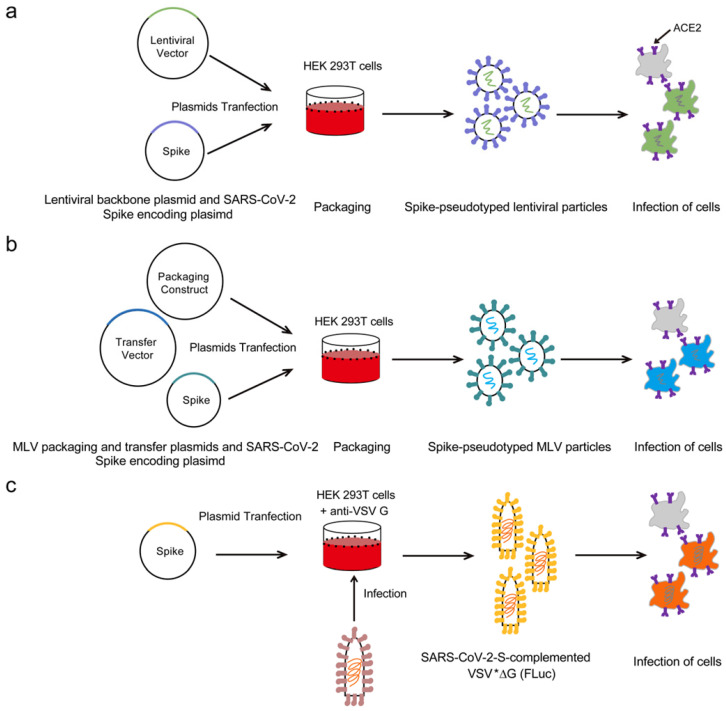
Pseudoviruses are exactly what they sound like: fake (fake) “viruses.” These are said to be recombinant “viruses” (i.e. lab-created cultured concoctions from many sources) which mix and match genetic material from other “viruses” making them “less virulent.” This is how pseudoviruses are described according to the study Construction and applications of SARS-CoV-2 pseudoviruses: a mini review:
“Pseudoviruses are a kind of recombinant virus with their core or backbone and surface proteins derived from different viruses9. Genes inside pseudoviruses are usually altered or modified to abolish native surface protein expression. An additional plasmid is then used to express alternative surface proteins, producing a pseudovirus that can infect susceptible host cells but can only replicate intracellularly for a single round10, 11. As viral surface proteins play pivotal roles in gaining entry into host cells, the conformational structures of pseudoviral surface proteins have high similarity to that of the native viral proteins; however, pseudoviruses have attenuated virulence compared with wild-type (WT) viruses, allowing them to be safely handled in biosafety level 2 laboratories.”
https://www.ncbi.nlm.nih.gov/pmc/articles/PMC8071765/figure/F1/?report=objectonly
Sadly, the researchers of the paper Hammond cited do not share how their pseudovirus was created, but from the above source we can obtain an idea of what was done through the cell culture process. The reseachers also claim that their findings using the fake (fake) “virus” need to be confirmed using the real (fake) “SARS-COV-2 virus.” Thus, once again, we can see that the study cited does not reflect reality in any way. Any and all conclusions about how the spike protein of “SARS-COV-2” would affect the vascular system can not be gained from this paper. All that can be taken away is that when mixing and matching different cultures, different indirect chemical results are generated outside of any living organism in a lab which can then be interpreted by the researchers into a story of how a fictional entity they never studied may act or behave while within the human body. Then the headlines and snippets from the conclusions can be used by bloggers and “fact-checkers” to bolster a fraudulent case claiming that part of the fictional entity is pathogenic by itself:
SARS-CoV-2 Spike Protein Impairs Endothelial Function via Downregulation of ACE 2
“We administered a pseudovirus expressing S protein (Pseu-Spike) to Syrian hamsters intratracheally. Lung damage was apparent in animals receiving Pseu-Spike, revealed by thickening of the alveolar septa and increased infiltration of mononuclear cells (Figure [A]).”
“Although the use of a noninfectious pseudovirus is a limitation to this study, our data reveals that S protein alone can damage endothelium, manifested by impaired mitochondrial function and eNOS activity but increased glycolysis. It appears that S protein in ECs increases redox stress which may lead to AMPK deactivation, MDM2 upregulation, and ultimately ACE2 destabilization.4 Although these findings need to be confirmed with the SARS-CoV-2 virus in the future study, it seems paradoxical that ACE2 reduction by S protein would decrease the virus infectivity, thereby protecting endothelium. However, a dysregulated renin-angiotensin system due to ACE2 reduction may exacerbate endothelial dysfunction, leading to endotheliitis. Collectively, our results suggest that the S protein-exerted EC damage overrides the decreased virus infectivity. This conclusion suggests that vaccination-generated antibody and/or exogenous antibody against S protein not only protects the host from SARS-CoV-2 infectivity but also inhibits S protein-imposed endothelial injury.”
https://www.ahajournals.org/doi/10.1161/CIRCRESAHA.121.318902
“A study in The FASEB Journal in May 2021 found that the spike protein induces acute lung injury in mice genetically engineered to express the ACE2 receptor, which is the receptor on certain human cells that enables the coronavirus via the spike protein to fuse with and enter the cells where it then replicates.”
Sadly, this next study remains blocked to me as I am unable to access the full paper. However, Hammond did supply a link which discussed the findings and from that article we can find out some interesting details:
“Studying SARS-CoV-2 can be challenging because experiments involving the intact virus requires a Biosafety Level 3 laboratory. To overcome this hurdle, the researchers created a new model of acute lung injury that utilizes transgenic mice that express the human receptor for SARS-CoV-2 in their lungs.
“Our mouse model dramatically reduces the danger of doing this type of research by allowing COVID-19 lung injury to be studied without using the intact, live virus,” said Solopov. “This will greatly increase and diversify the ability to do COVID-19 research. Our model will also likely be useful for studying other coronaviruses.”
The researchers injected the genetically modified mice with a segment of the spike protein and analyzed their response 72 hours later. Another group of mice received only saline to serve as a control.
The researchers found that the genetically modified mice injected with the spike protein exhibited COVID-19-like symptoms that included severe inflammation, an influx of white blood cells into their lungs and evidence of a cytokine storm – an immune response in which the body starts to attack its own cells and tissues rather than just fighting off the virus. The mice that only received saline remained normal.”
What we can see is that transgenic mice were used in order to study the so-called spike protein. Transgenic mice are genetically-altered mice which are said to have been constructed by injecting cloned DNA into fertilized mouse eggs. The eggs that survive this process are then implanted into female mice in order to develop them. These transgenic mice were then said to be injected with the spike protein into their throats. While I could not find any details about what spike protein was used in this study, it is reasonable to assume that this was yet another recombinant lab-created cultured protein as the researchers admitted to not using “live infectious virus” nor needing a level 3 biosafety laboratory. The transgenic mice came down with “COVID-like” symptoms which consisted of severe inflammation, increased white blood cell counts in the lungs, and “evidence” of a cytokine storm (which was only mentioned in the article yet not in the abstract nor conclusion of the study).
Thus, we have a study utlizing genetically-altered mice that experienced inflammation when injected unnaturally in the throat with lab-created cultured recombinant goo. From these results, the researchers confidentally proposed that a single exposure of K18-hACE2 mice to “SARS-CoV-2” Spike Protein S subunit S1 may represent a valid model of “COVID-19” that may be useful for the preclinical investigation of new potential countermeasures against “COVID-19” and other “coronaviral” infections. Hardly a ringing endorsement, which is not surprising given the experimental set-up, yet this did not stop Hammond from including it as part of his litany of “favorable” evidence:
Single intratracheal exposure to SARS-CoV-2 S1 spike protein induces acute lung injury in K18-hACE2 transgenic mice
“The SARS-CoV-2 pandemic has infected more than 85,900,000 people and provoked the death of more than 1.9 million worldwide. Therapeutic options remain limited, and vaccines may exhibit narrow efficacy, due to short supplies, delays in distribution and the emergence of new resistant strains. It is mandatory to study new therapeutic approaches that modulate the strong inflammatory response observed in the lung, prevent respiratory failure and improve outcomes. The study of SARS-CoV-2 pathogenicity in vivo is challenging due to the necessary biosafety laboratory regulations. Thus, we developed an acute lung injury model by intratracheally instilling the S1 subunit of SARS-CoV-2 Spike S protein (400 µg/kg, 2 ml/kg body weight) in K18-hACE2 transgenic mice that overexpress the human receptor for SARS-CoV-2 Spike protein S, ACE2, and investigated outcomes 72 hours later. Mice exhibited an acute decline in body weight during the first 48 hours following instillation, compared to saline-instilled controls. At 72 hours, bronchoalveolar lavage fluid demonstrated a dramatic increase in white blood cell content, particularly neutrophils, and marked proteinosis compared to controls. Histologic examination of lung tissue revealed hyaline membranes, alveolar septal thickening, and a large number of neutrophils in the interstitial and alveolar spaces of Spike protein S exposed mice. We propose that a single exposure of K18-hACE2 mice to SARS-CoV-2 Spike Protein S subunit S1 may represent a valid model of COVID-19, allow the study of the molecular mechanisms of SARS-CoV-2 induced lung injury and be useful in the investigation of potential new therapeutic approaches to the management of COVID-19 as well as future coronavirus-dependent respiratory diseases.
Conclusions: We demonstrate for the first time that the intratracheal instillation of a single element of the SARS-CoV-2 virus, the subunit 1 of the Spike protein, in K18-hACE2 transgenic mice, is capable of eliciting strong pulmonary and systemic inflammation, including activation of the STAT3 and NFκB pathways in the lungs and biochemical and histological evidence of acute lung injury. We propose that this model may be useful for the preclinical investigation of new potential countermeasures against COVID-19 and other coronaviral infections.”

https://doi.org/10.1096/fasebj.2021.35.S1.04183
“A study in Clinical Infectious Diseases in May 2021 showed that the spike protein induced by Moderna’s mRNA COVID‑19 vaccine circulates throughout the body.”
The problem with Hammond’s next study relates once again to the use of recombinant lab-created spike proteins as well as the use of serological results to conclude anything meaningful from the experimental procedures. This study attempts to claim that antibody results show that the spike protein, said to be created during the unobservable process after vaccination, circulates throughout the body. The researchers used different assays during the study and commented on the fact that no one had ever shown these kinds of results before. They admitted that this may have to do with the limitations of the assays themselves, thus showcasing the potential inaccuracy of their own results due to technological limitations.
Beyond the technological limitations is the problem of claiming that theoretical antibodies can detect theoretical spike proteins. In order for this to be true, the spike protein and the antibodies themselves must both be purified and isolated first and then experimented with together in order to show that they are specific to each other. As neither the spike protein nor the antibodies have ever been scientifically proven to exist in purified/isolated form, these results are essentially meaningless. One can not use one fictional entity to determine the presence and/or absence of another fictional entity. This is why non-specific results regularly occur with antibodies and we end up seeing statements from researchers such as “possibly due to assay cross-reactivity with other human coronaviruses” as we see in this study.
In any case, only 3 of 13 participants saw spike protein antibody results for 15 days after the first injection and none saw any spike protein antibody results for upwards of 56 days after the second injection. If the spike protein is supposed to circulate in the body after injection, based on these results, it doesn’t appear to do so for very long if at all in the vast majority of the participants studied:
Circulating Severe Acute Respiratory Syndrome Coronavirus 2 (SARS-CoV-2) Vaccine Antigen Detected in the Plasma of mRNA-1273 Vaccine Recipients
“Here we provide evidence that circulating SARS-CoV-2 proteins are present in the plasma of participants vaccinated with the mRNA-1273 vaccine. We report antigen and serological data of the mRNA-1273 vaccine in 13 healthcare workers at the Brigham and Women’s Hospital. Ultrasensitive single-molecule array (Simoa) assays were used for the detection of SARS-CoV-2 antigens spike (S1–S2 unit), S1, and nucleocapsid and antibodies immunoglobulin G (IgG), immunoglobulin A (IgA), and immunoglobulin M (IgM) against SARS-CoV-2 spike, S1, receptor binding domain (RBD), and nucleocapsid, as previously described [6, 7].”
“S1 antigen was detected as early as day 1 postvaccination, and peak levels were detected on average 5 days after the first injection (Figure 1A). The mean S1 peak level was 68 pg/mL ± 21 pg/mL. S1 in all participants declined and became undetectable by day 14. No antigen was detected at day zero for 12 of 13 participants, as expected. However, one individual presented detectable S1 on day zero, possibly due to assay cross-reactivity with other human coronaviruses or asymptomatic infection at the time of vaccination. Spike protein was detectable in 3 of 13 participants an average of 15 days after the first injection. The mean spike peak level was 62 pg/mL ± 13 pg/mL. After the second vaccine dose, no S1 or spike was detectable, and both antigens remained undetectable through day 56. For one individual (participant 8), spike was detected at day 29, 1 day after the second injection and was undetectable 2 days later.”
“We observe an increase in S1 over an initial period of 1–5 days, suggesting that mRNA translation begins immediately after vaccine inoculation. Interestingly, spike protein appears in 3 of 13 participants on average 8 days after S1 is produced. The Simoa antigen assays for the full spike protein are designed to require antibody binding to both the S1 and S2 subunits for detection, resulting in a cleaved spike protein to be undetectable. Additionally, spike protein concentrations in plasma of vaccinated participants may be below our assay limit of detection. We hypothesize that the cellular immune responses triggered by T-cell activation, which would occur days after the vaccination, lead to direct killing of cells presenting spike protein, and an additional release of spike into the blood stream [9]. The mechanisms underlying release of free S1 and the subsequent detection of the intact spike protein remain unclear and require further studies.”
“Limitations of the current study include the small sample size and potential biases that result from enrolling healthy, young adults, which may not be representative of the general population. Future studies should also examine the dynamics of antigen production with neutralization antibodies. Nonetheless, evidence of systemic detection of spike and S1 protein production from the mRNA-1273 vaccine is significant and has not yet been described in any vaccine study, likely due to limitations in assay sensitivity and timing assessment. The clinical relevance of this finding is unknown and should be further explored.”
From the supplemental material:
“SARS-CoV-2 Spike protein (produced in Bing Chen’s lab), Nucleocapsid recombinant protein (Ray Biotech 230-30164), S1 protein (Sino Biological 40591-V08H), and RBD (produced in Aaron Schmidt’s lab) were conjugated to 647 nm, 488 nm, 700 nm, and 750 nm dye-encoded carboxylated paramagnetic beads (Quanterix), respectively, using EDC (1-ethyl-3-(3-dimethylaminopropyl) carbodiimide hydrochloride) chemistry (ThermoFisher Scientific 77149).”
https://academic.oup.com/cid/article/74/4/715/6279075?login=false
“The authors of a paper published on the preprint website Authorea in May 2021 expressed concern that government authorities were minimizing or ignoring concerns about the potential toxicity and pathogenicity of the spike protein induced by vaccination. (A preprint study is one that has not yet undergone peer review.)”
This next paper is more of a commentary than an actual study. While the authors are right to suggest that the adverse vaccine reactions that occur can and will be counted as cases of “SARS-COV-2,” the evidence used to claim that the spike protein is pathogenic and the potential cause is based on Hammond’s earlier fraudulent study which used pseudoviruses and hamsters. Again, I am not stating that the vaccines are not dangerous. They most definitely are dangerous and toxic. However, the story that there is a pathogenic spike protein created from the mRNA injection is nothing but pure science fiction based upon fraudulent evidence that is circulated by those aiming to keep the confused public believing in the germ theory lie. Ironically, the authors end their commentary by saying that “relying on a careful evaluation of the relevant scientific research, is urgent” and that it “is imperative to follow the science.” Sadly, it appears that they did no such thing:
SARS-CoV-2 mass vaccination: Urgent questions on vaccine safety that demand answers from international health agencies, regulatory authorities, governments and vaccine developers
“Furthermore, even in the absence of SARS-CoV-2 virus, Spike glycoprotein alone causes endothelial damage and hypertension in vitro and in vivo in Syrian hamsters by down-regulating angiotensin-converting enzyme 2 (ACE2) and impairing mitochondrial function [26]. Although these findings need to be confirmed in humans, the implications of this finding are staggering, as all vaccines authorized for emergency use are based on the delivery or induction of Spike glycoprotein synthesis. In the case of mRNA vaccines and adenovirus-vectorized vaccines, not a single study has examined the duration of Spike production in humans following vaccination. Under the cautionary principle, it is parsimonious to consider vaccine-induced Spike synthesis could cause clinical signs of severe COVID-19, and erroneously be counted as new cases of SARS-CoV-2 infections. If so, the true adverse effects of the current global vaccination strategy may never be recognized unless studies specifically examine this question. There is already non-causal evidence of temporary or sustained increases in COVID-19 deaths following vaccination in some countries (Fig. 1) and in light of Spike’s pathogenicity, these deaths must be studied in depth to determine whether they are related to vaccination.”
“An open scientific dialogue is urgent and indispensable to avoid erosion of public confidence in science and public health and to ensure that the WHO and national health authorities protect the interests of humanity during the current pandemic. Returning public health policy to evidence-based medicine, relying on a careful evaluation of the relevant scientific research, is urgent. It is imperative to follow the science.”
“Ironically, Dr. Peter Hotez, whom Health Feedback cites to support its argument that the spike protein induced by vaccination is harmless, was among the authors of a study published in the journal Circulation in July 2021 noting that mRNA COVID‑19 vaccines can cause myocarditis, or inflammation of the heart, and hypothesizing that this might be due to some individuals’ immune response to either vaccine mRNA or the vaccine-induced spike protein.”
Hammond next supplied a review article by Dr. Peter Hotez which he used as evidence that the mRNA vaccine can cause myocarditis. In the review, Dr. Hotez and the other authors hypothesized (an educated guess) that myocarditis could be due to the spike protein. However, in the paper it was admitted that the mechanisms for the development of myocarditis were unknown and several possible proposals were listed. The study that was cited in the review which was used as evidence for the hypothesized role that the spike protein plays in myocarditis, used recombinant spike protein and commercially available antibodies in order to obtain the results. Once again, we have hypothetical conclusions crafted around results from lab-created concoctions which have no bearing on reality:
“Although the mechanisms for development of myocarditis are not clear, molecular mimicry between the spike protein of severe acute respiratory syndrome coronavirus-2 (SARS-CoV-2) and self-antigens, trigger of preexisting dysregulated immune pathways in certain individuals, immune response to mRNA, and activation of immunologic pathways, and dysregulated cytokine expression have been proposed.”
“Another important potential mechanism for myocarditis is molecular mimicry between the spike protein of SARS-CoV-2 and self-antigens.50 Antibodies against SARS-CoV-2 spike glycoproteins have been experimentally shown to cross-react with structurally similar human peptide protein sequences, including α-myosin.50 However, severe adverse events or autoimmune reactions have been very rare.46,47 Although COVID-19 vaccination does not appear to provoke de novo immune-mediated adverse events, it is possible that it may trigger preexisting dysregulated pathways in certain individuals with predisposition, resulting in a polyclonal B-cell expansion, immune complex formation, and inflammation.48″
https://www.ahajournals.org/doi/10.1161/CIRCULATIONAHA.121.056135
Link 50 takes us to this study:
Potential antigenic cross-reactivity between SARS-CoV-2 and human tissue with a possible link to an increase in autoimmune diseases
“Commercially available mouse monoclonal antibody made against recombinant SARS coronavirus spike protein and rabbit monoclonal antibody made against SARS coronavirus nucleoprotein were applied at optimal dilution to the SARS-CoV-2 proteins and to 50 different tissue antigens using enzyme-linked immunosorbent assay (ELISA). Recombinant SARS-CoV-2 spike protein S1 and recombinant SARS-CoV-2 nucleocapsid protein were purchased from RayBiotech. ELISA wells were coated with nuclear antigens, dsDNA, F-actin, and mitochondria (M2) antigen purchased from different companies. An additional 45 tissue antigens used in this study have been previously described [9].”
https://www.sciencedirect.com/science/article/pii/S1521661620304253?via%3Dihub
“A study published at the preprint server bioRxiv in July 2021 found an association between “Long Covid” and persistence of the spike protein in the absence of persistence of whole viable virus, once again indicating that the spike protein alone is pathogenic.”
This next study provided by Hammond tried to make the case that detecting the spike protein RNA without finding whole “SARS-COV-2” RNA/genome was sufficient to claim that the spike protein itself was pathogenic and persisting within the body. To deternine this, the researchers first used digital droplet PCR to find “SARS-COV-2” RNA in the blood of a few patients, thus relying on a fraudulent test and genome to determine the initial findings. This step resulted in 36% (4 of 11) of severe “COVID-19” patients’ PBMCs contained “SARS-CoV-2” RNA compared to 4% (1/26) of PASC patients’ PBMCs. The researchers then decided to use flow cytometry with antibodies said to define B cell, T-cell, and monocytic subsets in addition to simultaneous staining of these cells with an antibody claimed to be specific to the “SARS-CoV-2” S1 protein to try and determine the reservoir for the detected RNA. To confirm the presence of “SARS-CoV-2” S1 protein, they then sorted CD14lo, CD16+ monocytes and performed Ultra High-Performance Liquid Chromatography. After various preparation processes, the researchers measured the peptides coming from peripheral blood monolayers (stored in 90% fetal bovine serum) and compared them to a peptide database from a commercially made recombinant spike protein and found up to 44% (i.e. up to = not always that high) of the S1 subunit peptides could be identified in patient samples. In other words, the researchers used indirect measurements of peptides matched between patient samples and recombinant spike proteins which were mapped to a peptide database created from the commercially made recombinant protein in order to claim INDIRECTLY that the spike protein was in the patients samples. Can you spot the circular methods there?
Worse yet, full length sequencing of the five cases submitted for genomic analysis failed to identify any full-length sequence in the spike protein gene, or any other gene, that could account for the observed spike protein detected by proteomic analysis. All they could find were fragments of “SARS-COV-2” genomes. Thus, they concluded that it must not be replication competent “virus” that was found but non-infectious leftover spike proteins which were not cleared from the body. As can be seen, this smattering of indirect evidence is based upon a “virus” and spike protein that was never properly purified and isolated first in order to accurately determine any specific antibody responses nor determine an accurate genome and peptide database. Thus, all measurements used to indirectly claim the presence of the spike protein are based on results coming from fraudulent lab-generated cultured creations:
Persistence of SARS CoV-2 S1 Protein in CD16+ Monocytes in Post-Acute Sequelae of COVID-19 (PASC) Up to 15 Months Post-Infection
“Since the reports by our group and others found that monocyte subsets can be infected by HIV, HCV, Zika virus and Dengue fever virus (10–12), we screened peripheral blood mononuclear cells (PBMCs) from PASC individuals, as well as acute severe COVID-19 as controls, for SARS-CoV-2 RNA (Table 1). Using the highly sensitive, quantitative digital droplet PCR (ddPCR), we found that 36% (4 of 11) of severe COVID-19 patients’ PBMCs contained SARS-CoV-2 RNA compared to 4% (1/26) of PASC patients’ PBMCs. The one PASC patient that was RNA positive was 15 months post infection.
To further establish the exact reservoir contributing to the positive signal detected using ddPCR, we performed high parameter flow cytometry with antibodies that define B cell, T-cell, and monocytic subsets in addition to simultaneous staining of these cells with an antibody for the SARS-CoV-2 S1 protein. As demonstrated in Figure 2, we found distinct subpopulations of SARS-CoV-2 containing cells in the CD14lo, CD16+ monocytic subset for 73% (19 out of 26) of PASC patients and 91% (10 out of 11) of severe COVID-19 patients. As demonstrated in Figure 3, the quantity of SARS-CoV-2 S1 containing cells were statistically significant in both the severe patients (P=0.004) and in the PASC patients (P=0.02). Neither classical monocytes nor intermediate monocytes expressed the SARS-CoV-2 S1 protein.
To confirm the presence of SARS-CoV-2 S1 protein, we sorted CD14lo, CD16+ monocytes and performed Ultra High-Performance Liquid Chromatography (UHPLC). Following immunoprecipitation, the elution fractions were dried down in vacuo, resuspended in ddH2O and purified by to remove any non-crosslinked SARS-CoV-2 S1 antibody as well as any detergents from the commercial immunoprecipitation buffers. The UHPLC collected fractions were dried in vacuo, resuspended in 100 mM HEPES (pH 8.0, 20% Acetonitrile), and subjected to cistern: reduction and alkylation with chloroacetamide. The samples were then digested with AspN and LysC endopeptidases for 16h at 37°C. The digested peptides were analyzed on an Agilent 6550 IonFunnel QTOF and 1290 UHPLC by comparing patient samples to identical digests performed on commercially available SARS-CoV-2 S1 subunit. S1 subunit peptides from patient samples were mapped to a peptide database generated using commercial S1 subunit digests. Peptide identification consisted of matches in exact mass, isotope distribution, peptide charge state, and UHPLC retention time. As shown in Figure 4, the retention time of the representative peptide NLREFVFK in the digested commercial S1 subunit and Sample LH1-6 matched. Additionally, the Mass Spectra in Figure 4 show identical mass, isotope distribution, and charge states for the representative peptide NLREFVFK in the representative LH1 sample and commercial S1 subunit (also observed in LH 2-6, not shown). Using these metrics, up to 44% of the S1 subunit peptides could be identified in patient samples LH1-LH6 (Supplementary Table 1), providing complementary evidence to flow cytometry experiments that demonstrate the presence of S1 subunit protein in these patient cells.”
“Cells from 4 out of 11 severe COVID-19 patients and 1 out of 26 PASC patients contained ddPCR+ peripheral blood mononuclear cells, however, only fragmented SARS-CoV-2 RNA was found in PASC patients. No full length sequences were identified, and no sequences that could account for the observed S1 protein were identified in any patient.”
“It is important to note that the S1 protein detected in these patients appears to be retained from prior infection or phagocytosis of infected cells undergoing apoptosis and is not the result of persistent viral replication. Full length sequencing of the five cases submitted for genomic analysis failed to identify any full-length sequence in the spike protein gene, or any other gene, that could account for the observed spike protein detected by proteomic analysis.”
High Parameter Immune Profiling/Flow Cytometry
Peripheral blood mononuclear cells were isolated from peripheral blood using Lymphoprep density gradient (STEMCELL Technologies, Vancouver, Canada). Aliquots 200 of cells were frozen in media that contained 90% fetal bovine serum (HyClone, Logan, UT) and 10% dimethyl sulfoxide (Sigma-Aldrich, St. Louis, MO) and stored at -70°C. Cells were stained and analyzed using a 17-color antibody cocktail including a PE-labeled SARS-CoV-2 S1 antibody (BioTechne, Minneapolis MN).
LC-MS analysis
Digested recombinant SARS-CoV-2 Spike S1 protein was analyzed by a high mass accuracy mass spectrometer to generate a list of detectable peptides with retention time and accurate masses. An Agilent 1290 Infinity II high pressure liquid chromatography (HPLC) system and an AdvanceBio Peptide Mapping column (2.1 × 150 mm, 2.7 μm) were used for peptide separation prior to mass analysis.”
https://www.biorxiv.org/content/10.1101/2021.06.25.449905v3.article-info
“A study in Viruses in October 2021 found that the spike protein can enter the nucleus of cells and inhibit DNA damage repair. The authors noted that this finding was relevant for mRNA COVID‑19 vaccines since they are designed to elicit human cells to produce the spike protein.”
Interestingly, this next source supplied by Hammond to state that the spike protein can enter the nucleus of cells and inhibit DNA damage repair was actually retracted May 10th, 2022, well over a month before his article was published on June 28th, 2022.
Retraction published on 10 May 2022, see Viruses 2022, 14(5), 1011.
This is a perfect example as to why one should never just read the headline and throw any study out there in support of an argument without actuallly having read the study first. All claims from this paper which were used by Hammond in support of his spike protein argument are therefore false and invalid:
SARS–CoV–2 Spike Impairs DNA Damage Repair and Inhibits V(D)J Recombination In Vitro
“The published article [1] has been retracted. Following publication, the first author contacted the editorial office regarding an improper experimental design with the potential to significantly affect the integrity of the resultant experimental data.
Adhering to our complaint procedure, an investigation was conducted. Both the chosen construct of the spike plasmid that contained a C-terminal fused with 6xHis tag and use of a GFP reporter system under overexpression conditions in the protocol were identified as having the potential to introduce significant ambiguity regarding the nature of the reported observations. The reliability of the results and conclusions presented have therefore been undermined. Furthermore, statements regarding the effect of the spike protein on the adaptive immunity are misleading as in this article no experiments related to the adaptive immunity were performed, and the full-length spike-based vaccine was not studied. Therefore, conclusions related to vaccine safety are not validated and lacked experimental support. This article [1] is retracted and shall be marked accordingly. This retraction was approved by the Editor-in-Chief of the journal Viruses.”
https://www.mdpi.com/1999-4915/13/10/2056/htm
“A study in the Journal of Immunology in November 2021 found that the spike proteins induced by the Pfizer-BioNTech COVID‑19 vaccine are carried by extracellular vesicles called exosomes and circulate throughout the body, which helps to explain the blood antibody response elicited by vaccination.”
In Hammond’s next study, it is claimed that the vaccines create exosomes which carry spike proteins on their surface that circulate throughout the body. This is yet another serological study attempting to claim that the antibodies used are specific to a spike protein which was used in order to stain the exosome so that the spike proteins can be visualized. However, once again it must be stated that in order to deternine specific antibody responses, the “virus,” it’s spike protein, the antibody, and the exosomes would have needed to have been properly purified and isolated directly from the fluids of humans first. This has never been done and thus these all remain unproven fictitious entities which can not be used in order to verify the existence of the other. This is yet another study using recombinant cultured goo said to contain spike proteins and not purified/isolated particles proven to be spike proteins. It all amounts to nothing more than staining fluids and pointing and declaring at the dots which stick to a blob in EM and claiming this as proof that exosomes with spike proteins were created from vaccination:
Cutting Edge: Circulating Exosomes with COVID Spike Protein Are Induced by BNT162b2 (Pfizer–BioNTech) Vaccination prior to Development of Antibodies: A Novel Mechanism for Immune Activation by mRNA Vaccines
“In the current study, we analyzed eight healthy adults who received both doses of the SARS-CoV-2 vaccine (Pfizer–BioNTech). Our results demonstrated the induction of circulating exosomes carrying the SARS-CoV-2 spike protein by day 14, when Abs to the spike protein were not detectable in the sera using an ELISA method developed in our laboratory. Circulating Abs were detectable only after the second booster dose of vaccine (days 14), and the amount of exosomes containing spike protein was increased up to ∼12-fold maximum.”
Materials and Methods
Patient cohort and demographics
We analyzed eight healthy adult volunteers vaccinated with the mRNA-based SARS-CoV-2 vaccine (Pfizer–BioNTech). Blood was collected before vaccination, days 7 and 14 after the first dose, day 14 after the second dose, and 4 mo after both the doses. This study was approved by the Institutional Review Boards (IRB) at St. Joseph’s Hospital (IRB number PHXB16-0027-10-18).
Exosome isolation and nanoparticle tracking analysis
Exosomes were isolated from 500 µl of plasma using Invitrogen Exosome Isolation Kit followed by 0.22-micron filtration (7). All exosomes were analyzed for size by NanoSight NS300 (Malvern Panalytical, Great Malvern, U.K.), and the mean size of the particles used in our experiments was <200 nm (8).
Detection of Abs to SARS-CoV-2 spike protein and nucleocapsid protein from human plasma samples
Development of Abs to SARS-CoV-2 spike Ag was determined using an ELISA developed in our laboratory. In brief, 1 μg/ml SARS-CoV-2 spike protein (Sino Biological) suspended in PBS was coated on an ELISA plate and incubated overnight at 4°C. Human plasma was added to these plates at 1:750 dilution. Detection was performed using secondary anti-human IgG-HRP (1:10,000) and developed using tetramethylbenzidine substrate and read at 450 nm.”
Transmission electron microscopy of isolated exosomes for SARS-CoV-2 spike protein
“Exosomes were labeled with immunogold and mouse anti–SARS-CoV-2 spike Ab, and coronavirus FIPV3-70 Ab (1:100) was added to the grids. Grids were washed and stained with uranyl acetate and viewed by transmission electron microscopy (JEOL USA, Peabody, MA) (10).”
“We performed transmission electron microscopy using Abs specific for SARS-CoV-2 spike to demonstrate the presence of SARS-CoV-2 Ags on the surface of exosomes from controls and healthy vaccinated individuals. Exosomes from vaccinated individuals are positive for SARS-CoV-2 Ag (Fig. 1B). We have also stained both the exosome samples with coronavirus FIPV3-70 Ab as negative control and did not observe any positive reaction in exosomes (Fig. 1B).”

https://www.jimmunol.org/content/207/10/2405#ref-7
“A study in Clinical Science in December 2021 tested the hypothesis that the spike protein “may act as a ligand to induce non-infective cellular stress”. They showed that exposure to the spike protein alone “elicited signalling and functional alterations”, including “secretion of pro-inflammatory molecules typically involved in the cytokine storm” and “production of pro-apoptotic factors” causing endothelial cell death.
Health Feedback briefly notes this study by saying that it “showed that the spike protein can affect heart cells in the lab, but they used higher amounts of the protein than those found in COVID‑19 patients.”
Nevertheless, the study did demonstrate a biologically plausible mechanism by which blood clots could occur with SARS‑CoV‑2 infection or vaccination.”
This was another study used by Hammond to try and claim that the spike protein alone is pathogenic. He believed that the study showed a plausible way blood clots may occur due to “SARS-COV-2” and/or injection from the mRNA vaccine. However, once again, this study did not use purified and isolated particles but instead experimented with lab-created recombinant S proteins made from insect cells. The primary cell cultures used for the study were grown in dedicated medium supplemented with human recombinant growth factors and 2% fetal calf serum which hardly sounds like something the cells would encounter within a living organism. These cells were passaged between 4 to 7 times which can have detrimental effects on the culture as the passage number increases. The cell line cultures consisted of human gut epithelial cell line, Caco2, expressing hACE2 as well as African green monkey kidney cell line VeroE6 engineered to overexpress the human ACE2 and TMPRSS2. All cells were cultured in Dulbecco’s modified Eagle’s medium plus GlutaMAX supplemented with 10% FBS, 1% sodium pyruvate, and 0.1 mM non-essential amino acids. The human lung epithelial cell line Calu3 (ATCC HTB-55) was cultured in Eagle’s minimum essential medium plus GlutaMAX with 10% FBS, 0.1 mM non-essential amino acids, and 1% sodium pyruvate. In other words, there is absolutely nothing natural about the materials nor the chemical additives that they were kept in and experimented with.
The researchers stated that their study provided novel (as in fictional) proof-of-concept evidence for S protein capacity to cause molecular and functional changes in human vascular PCs. However, they admitted that their small sample size was inadequate and that further investigation in a larger population of patients was warranted to determine the cause for the inter-individual variability in PC infection. They also could not exclude that different scenarios may happen in vivo, i.e. within a living organism, as compared to that seen in vitro, i.e. inside a petri dish in a lab, thus essentially admitting that their results can not be applied to what occurs within a human body. Interestingly, the researchers also admitted that low amounts of the S protein could be detected in pre-pandemic control sera. They stated that this could be explained by the sequence homology between some regions of the S protein and other human proteins/peptides. In other words, the S protein contains similar sequences to normal human proteins/peptides and thus the tests that the researchers were using may have been picking up nothing more than normal human proteins/peptides rather than the theoretical S protein. Sadly, the immunogen sequence for the ELISA kit they used was locked away behind proprietary information (as is always the case), and therefore they could not determine if it recognised the S protein residues that have homology with unrelated peptides. Thus, the results from this study truly were worthless:
The SARS-CoV-2 Spike protein disrupts human cardiac pericytes function through CD147 receptor-mediated signalling: a potential non-infective mechanism of COVID-19 microvascular disease
“Exposure to the recombinant S protein alone elicited signalling and functional alterations, including: (1) increased migration, (2) reduced ability to support endothelial cell (EC) network formation on Matrigel, (3) secretion of pro-inflammatory molecules typically involved in the cytokine storm, and (4) production of pro-apoptotic factors causing EC death.”
Primary cell cultures
“Cardiac PCs were immunosorted as CD31neg/CD34pos cells from human myocardial samples, and expanded in a dedicated medium supplemented with human recombinant growth factors and 2% v/v foetal calf serum (FCS) (ECGM2 complete kit, C-22111, PromoCell) as previously described [11,28]. Briefly, samples were finely minced using scissors and scalpel until nearly homogenous and digested with Liberase (Roche) for up to 1 h at 37 C, with gentle rotation. The digest was passed through 70-, 40-, and 30-μm strainers. Finally, the cells were recovered and sorted using anti-CD31 and -CD34 microbeads (Miltenyi) to deplete the population of CD31pos ECs and select CD31neg/CD34pos cells, which distinguish a population of perivascular cells in situ [11,28]. After expansion to passage 3, the purity of the cell population was verified using immunocytochemistry (ICC) or flow cytometry [11,28].
Human coronary artery ECs (CAECs) were purchased from PromoCell and expanded in the same medium used for PCs. All cells used in the present study tested negative for mycoplasma contamination (assessed using the PCR Mycoplasma Test Kit I/C, PromoCell, cat# PK-CA91-1096). Cells were used between passages 4 and 7.
Cell line cultures
The human gut epithelial cell line, Caco2, expressing hACE2 (Caco-2-ACE2) was a kind gift from Dr Yohei Yamauchi, University of Bristol. The African green monkey kidney cell line VeroE6 engineered to overexpress the human ACE2 and TMPRSS2 (VeroE6/ACE2/TMPRSS2) [29] was a kind gift from Dr Suzannah Rihn, MRC-University of Glasgow Centre for Virus Research. All cells were cultured in Dulbecco’s modified Eagle’s medium plus GlutaMAX (DMEM, Gibco, Thermo Fisher, cat# 10567014) supplemented with 10% v/v FBS (Gibco, Thermo Fisher, A3840001), 1% v/v sodium pyruvate, and 0.1 mM non-essential amino acids. The human lung epithelial cell line Calu3 (ATCC HTB-55) was cultured in Eagle’s minimum essential medium plus GlutaMAX (MEM, Gibco, Thermo Fisher, cat# 41090036) with 10% v/v FBS, 0.1 mM non-essential amino acids, and 1% v/v sodium pyruvate.”
Measurement of S protein in patients’ sera
“The presence of S protein in COVID-19 patients’ serum was evaluated using the COVID-19 Spike Protein ELISA Kit from Abcam (ab274342), according to manufacturer’s instructions. Pre-pandemic sera were employed as controls. All test sera were diluted 1:2. The S protein concentration was expressed as nanogram per millilitre serum. The antibody supplied in the kit recognised the S2 domain.”
Production and purification of the recombinant SARS-CoV-2 S protein
“SARS-CoV-2 S protein was expressed in insect cells and purified as described previously [33,35]. Briefly, the S construct encoded amino acids 1–1213 (extracellular domain – ECD) fused with a thrombin cleavage site, followed by a T4-foldon trimerisation domain and a hexahistidine (HIS) affinity purification tag at the C-terminus. The polybasic furin cleavage site was mutated (RRAR to A) to increase the stability of the protein for in vitro studies [33,35]. S protein was expressed in Hi5 cells using the MultiBac system [36]. Secreted S protein was harvested 3 days after infection by centrifuging the cell culture at 1000×g for 10 min followed by another centrifugation of supernatant at 5000×g for 30 min. S protein-containing medium was incubated with HisPur Ni-NTA Superflow Agarose (Thermo Fisher Scientific) for 1 h at 4°C. Resin bound with S protein was separated from unbound proteins and medium using a gravity flow column, followed by 30 column volume wash with wash buffer (65 mM NaH2PO4, 300 mM NaCl, 20 mM imidazole, pH 7.5). Finally, the protein was eluted with a step-gradient of elution buffer (65 mM NaH2PO4, 300 mM NaCl, 235 mM imidazole, pH 7.5). Eluted fractions were analysed by reducing SDS/PAGE. Fractions containing the S protein were pooled and concentrated using 50-kDa MWCO Amicon centrifugal filter units (EMD Millipore). During concentration, proteins were buffer-exchanged in PBS, pH 7.5. Concentrated protein was aliquoted, flash-frozen in liquid nitrogen, and stored at −80°C until use. In all the in vitro experiments of the manuscript, we will refer to the S-ECD protein simply as S protein.
Recombinant Spike S1 (#10522-CV) and S2 (#10584-CV) were purchased from R&D, resuspended in PBS according to manufacturer’s instructions, aliquoted and stored at −80°C until use. Similarly to the S-ECD, the S1 and S2 proteins were produced in insect cells.”
Discussion
“Our study provides novel proof-of-concept evidence for S protein capacity to cause molecular and functional changes in human vascular PCs, either dependently or independently of the CD147 receptor (summarised in Figure 11).”
“Here, we report that two-third of patients tested did not have their PCs infected by SARS-CoV-2, while the rate of infection was below 8% in the remaining subjects, suggesting a very low permissiveness of these cells to the coronavirus, at least in vitro.”
“Further investigation in a larger population of patients is warranted to determine the cause for the inter-individual variability in PC infection. Moreover, we cannot exclude different scenarios may happen in vivo.”
“In our study, low amounts of the S protein could be detected in pre-pandemic control sera. This could be explained by the sequence homology between some regions of the S protein and other human proteins/peptides. A previous report identified pathogenic regions of SARS-CoV-1 S protein, which share sequence homology with Angrgm-52 (GenBank accession number AAL62340), a novel gene up-regulated in human mesangial cells stimulated by angiotensin II and bradykinin [53]. Unfortunately, the immunogen sequence for this particular ELISA kit ab274342 is proprietary information, therefore we could not determine if it can recognise the S protein residues that have homology with unrelated peptides.”
Study limitations
“The study was conducted on isolated cells and therefore the evidence must be confirmed in vivo.
The amount of S protein used for in vitro studies was higher than the average S protein concentration detected in COVID-19 patients’ serum. However, circulating S protein represents the spill-over from infected organs, where concentration may be higher due to retention at the receptor level. Because we do not have access to post-mortem myocardial samples, we could not verify this hypothesis.”
“A study at bioRxiv on December 2021 showed that the SARS‑CoV‑2 spike protein by itself promoted platelet activation, which the authors suggested was “a pathogenic mechanism to explain thrombosis associated to COVID‑19 lung disease, by which Spike present in SARS‑CoV‑2 virions or exposed on the surface of infected cells, leads to platelet stimulation and subsequent activation of the coagulation cascade.” (Emphasis added.)
That study was also cited by The Epoch Times, to which Health Feedback responded by noting that the researchers “studied the mechanisms for formation of blood clots, showing that spike protein can activate platelets in the lab but they did not establish if this happened at concentrations found in patients or vaccinated people.”
In this second to last study, Hammond relied on a preprint non-peer-reviewed paper to share the author’s suggestion that the spike protein can act alone in order to cause thrombosis in “Covid” patients. The paper comes with the below warning that the study should not be reported as conclusive:
A reminder: they have not been formally peer-reviewed and should not guide health-related behavior or be reported in the press as conclusive.
Interestingly, in a moment of shooting his own argument in the foot, Hammond pointed out that the Health Feedback article rightly stated that the results were only relevant in the lab and that the researchers did not establish if the results with the recombinant spike protein happened at concentrations found in patients or vaccinated people. This may have to do with the fact that, once again, the study relied on cell-cultured creations and “pseudoviruses” to generate their conclusions.
For some reason, I was unable to copy/paste the relevant sections from the study but I am providing the methods section from the abstract as well as a few images from the PDF showing that the researchers utilized cell cultured creations and lab-created “pseudoviruses” for their experiments. You can see the various processes used to create their fictional entity. Thus, once again, the experimental results do not reflect reality in any way:
SARS-CoV-2 Spike protein activates TMEM16F-mediated platelet pro-coagulant activity
“Methods We produced SARS-CoV-2 Spike or VSV-G protein-pseudotyped virions, or generated cells expressing Spike on their plasma membrane, and tested their effects on platelet adhesion (fluorescence), aggregation (absorbance), exposure of phosphatidylserine (flow cytometry for annexin V binding), calcium flux (flow cytometry for fluo-4 AM), and clot formation and retraction. These experiments were also conducted in the presence of the TMEM16F activity inhibitors Niclosamide and Clofazimine.”


https://www.biorxiv.org/content/10.1101/2021.12.14.472668v2
“A commentary in Clinical Science on March 29 of this year summarized the state of knowledge about the pathogenicity of the spike protein alone:”
Finally, we have made it to the last of Hammond’s exhausting list of pseudoscientific sources he seemingly failed to read. This one is a commentary on the state of “knowledge” on the lab-created recombinant and/or pseudovirus spike protein acting pathogenically alone. In other words, it added nothing new and actually used the same Avolio et al. study Hammond used which I outlined two sections above. If you can recall, this was the study which utilized lab-created recombinant S proteins made from insect cells for their experiments. The reseachers could not exclude that different scenarios may happen in vivo, i.e. within a living organism, as compared to that seen in vitro, i.e. inside a petri dish in a lab, thus essentially admitting that their results can not be applied to what occurs within a human body. They also admitted that the S protein contains similar sequences to normal human proteins/peptides and that the tests that the researchers were using may have been picking up nothing more than normal human proteins/peptides rather than the theoretical S protein. In other words, the evidence that the spike protein acts alone pathogenically from this commentary is as worthless as the Avolio et al. study from which it came:
“The concept that the S protein can cause detrimental effects in COVID-19 patients independent of infection could partially explain the long-term health issues.”
“There has been extensive evaluation of SARS-CoV-2 infection and COVID-19 on cardiovascular health [1,2]; however, there is emerging evidence that the S protein shed from SARS-CoV-2 can circulate in the blood of patients and have detrimental consequences [3,10,11]. (Figure 1) The study by Avolio et al. in this issue of Clinical Science provides strong evidence that the circulating S protein could be more detrimental to cardiac health than infection of SARS-CoV-2 to the heart [3]. More specifically, the S protein was found to act through the CD147 receptor on human cardiac pericytes to cause microvascular dysfunction [3]. The authors also determined that the S protein caused human cardiac pericyte inflammation via yet to be determined mechanisms [3].”
“The current study by Avolio et al. found similar results with the Alpha and Delta variants of the S protein on human cardiac pericytes [3]. Although the findings of the current study are provocative, future investigations need to expand the doses of S proteins to lower levels and other S protein variants for in vitro studies. Lastly, as we investigate SARS-CoV-2 and the circulating S protein and the impact on human health, we will be better prepared to treat patients as COVID-19 moves from a pandemic to an endemic status.”

In Summary:
- According to Jeremy Hammond, numerous studies have indicated that the spike protein of “SARS‑CoV‑2” by itself, in the absence of whole viable “virus,” can have pathogenic and toxic effects
- However, the studies supplied do not use purified/isolated spike proteins but recombinant proteins which are a manipulated form of protein, which is generated in various ways to produce large quantities of proteins, modify gene sequences and manufacture useful commercial products
- Recombinant protein is encoded by a gene — recombinant DNA — that has been cloned in a system that supports expression of the gene and translation of messenger RNA
- In such cases the system in which the protein is expressed must be easy to culture and maintain, grow rapidly, and produce large amounts of protein
- The various protein expression systems are:
- Bacteria
- Yeast
- Insect
- Mammalian
- A few of Hammond’s studies also relied on “pseudoviruses” which are a kind of recombinant “virus” with their core or backbone and surface proteins derived from different “viruses”
- Genes inside “pseudoviruses” are usually altered or modified to abolish native surface protein expression
- An additional plasmid is then used to express alternative surface proteins, producing a “pseudovirus” that can infect susceptible host cells but can only replicate intracellularly for a single round
- As “viral” surface proteins are said to play pivotal roles in gaining entry into host cells, the conformational structures of “pseudoviral” surface proteins have high similarity to that of the native “viral” proteins; however, “pseudoviruses” have attenuated virulence compared with wild-type (WT) “viruses,” allowing them to be safely handled in biosafety level 2 laboratories

- “SARS-CoV-2” subunit S1 (RayBiotech, Cat No 230–01101)
- “SARS-CoV-2” RBD (RayBiotech, Cat No 230–01102)
- “SARS-CoV-2” subunit S2 (RayBiotech, Cat No 230–01103)
- All three are recombinant “spike proteins” expressed in E. Coli and maintained in a filtered solution in 50 mM Na2HPO4 (pH 7.4), 0.5 M NaCl, 450 mM imidazole, and 6 M urea
- “SARS-CoV-2 S1” (RayBiotech, Cat No 230–30161-10)
- A recombinant “spike protein” grown in Human embryonic kidney 293 (HEK293) cells and supplied as a 0.2 um filtered solution in PBS (pH 7.4)
- “SARS-CoV-2 S2” also derived from HEK293 cells were used in experiments where indicated
- Other recombinant proteins (i.e TNFα) were purchased from R&D Systems (Minneapolis, MN, USA)
- To test functional outcomes, two (a 2D and a 3D vessel-like) in-vitro models of the blood brain barrier (BBB) using primary brain endothelial cells were used
- Primary human brain microvascular endothelial cells (hBMVECs) were isolated from fetal brain tissue
- Cells were grown on rat tail collagen I coated flasks (BD Biosciences) in full growth medium (EBM-2 medium supplemented with EGM-2MV SingleQuots (Lonza, Cat No CC-3156 and CC-4147)) in an incubator set to 37 °C, 5% CO2, and 100% humidity and experiments were performed in the basal medium (EBM2 supplemented with 10% FBS)
- These systems (when also coupled with other cells) represent the most advanced recapitulation (to reproduce or closely resemble) of the blood-brain barrier
- These results suggest that whether free “viral” spike proteins or those on the surface of the “virus” present during “SARS-CoV-2” infection could induce barrier permeability (albeit once a certain threshold is reached) equivocal to the concentrations used here (i.e. these results only relate to the lab-created conditions and concentrations used for the study)
- As this is the first report on the topic, they admit much work remains, particularly in regard to how permeability dynamics may change once these 3D microfluidic constructs are used with the whole “SARS-CoV-2 virus”

- It was found that the treatment of cultured primary human pulmonary artery smooth muscle cells (SMCs) or human pulmonary artery endothelial cells with the recombinant “SARS-CoV-2” spike protein S1 subunit is sufficient to promote cell signaling without the rest of the “viral” components
- In the cell culture experiments, two recombinant “SARS-CoV-2” spike proteins were studied
- Cultured primary human pulmonary artery SMCs and human pulmonary artery endothelial cells were treated with these proteins for 10 min
- They used the “phospho-specific” MEK antibody to determine that the recombinant full-length S1 subunit of “SARS-CoV-2” alone at a concentration as low as 130 pM, activated MEK
- The authors claim that recent observations suggest that the “SARS-CoV-2” spike protein can by itself trigger cell signaling that can lead to various biological processes
- Thus, they determined it is reasonable to assume that such events, in some cases, result in the pathogenesis of certain diseases
- According to the authors, this protein may also affect the cells of systemic and coronary vasculatures, eliciting other cardiovascular diseases such as coronary artery disease, systemic hypertension, and stroke (i.e. it is pure speculation as there is no evidence of this)
- In addition to cardiovascular cells, other cells that express ACE2 have the potential to be affected by the “SARS-CoV-2” spike protein, which may cause adverse pathological events (i.e. it is pure speculation as there is no evidence of this)
- The authors state that it is important to consider the possibility that the “SARS-CoV-2” spike protein produced by the new “COVID-19” vaccines triggers cell signaling events that promote PAH, other cardiovascular complications, and/or complications in other tissues/organs in certain individuals (i.e. it is pure speculation as there is no evidence of this)

- The researchers administered a pseudovirus expressing S protein (Pseu-Spike) to Syrian hamsters intratracheally (through the throat)
- Lung damage was apparent in animals receiving Pseu-Spike, (i.e. fake-spike…granted that’s an ironic admission as they all are) revealed by thickening of the alveolar septa and increased infiltration of mononuclear cells
- The researchers admitted that the use of a noninfectious pseudovirus is a limitation to this study
- However, they claim that their data revealed that (fake) S protein alone can damage endothelium, manifested by impaired mitochondrial function and eNOS activity but increased glycolysis
- It appeared that (fake) S protein in ECs increases redox stress which may lead to AMPK deactivation, MDM upregulation, and ultimately ACE2 destabilization
- They then admitted that their findings needed to be confirmed with the “SARS-CoV-2 virus” in the future study thus showing that their paper is utterly meaningless and irrelevant

- Studying “SARS-CoV-2” can be challenging because experiments involving the intact “virus” requires a Biosafety Level 3 laboratory
- To overcome this hurdle, the researchers created a new model of acute lung injury that utilizes transgenic mice (genetically-altered mice) that express the human receptor for “SARS-CoV-2” in their lungs
- “Our mouse model dramatically reduces the danger of doing this type of research by allowing COVID-19 lung injury to be studied without using the intact, live virus”
- The researchers injected the genetically modified mice with a segment of the spike protein and analyzed their response 72 hours later
- The genetically modified mice injected with the spike protein exhibited “COVID-19-like” symptoms that included:
- Severe inflammation
- An influx of white blood cells into their lungs
- Evidence of a cytokine storm
- Is injecting cultured goo down the throats of genetically altered mice not supposed to result in severe inflammation and increased WBC’s in the blood?
- Thus, they claim to have developed an acute lung injury model by intratracheally instilling the S1 subunit of “SARS-CoV-2” Spike S protein (400 µg/kg, 2 ml/kg body weight) in K18-hACE2 transgenic mice that overexpress the human receptor for “SARS-CoV-2” Spike protein S, ACE2, and investigated outcomes 72 hours later
- They proposed that a single exposure of K18-hACE2 mice to “SARS-CoV-2” Spike Protein S subunit S1 may represent a valid model of “COVID-19,” allow the study of the molecular mechanisms of “SARS-CoV-2” induced lung injury and be useful in the investigation of potential new therapeutic approaches to the management of “COVID-19” as well as future “coronavirus-dependent respiratory diseases”
- No mention of how the spike protein was created/aquired but it was most likely yet again a recombinant protein due to the fact that the researchers were not using “intact live viruses” nor in need of a biosafety level 3 facility

- Ultrasensitive single-molecule array (Simoa) assays were used for the detection of “SARS-CoV-2” antigens spike (S1–S2 unit), S1, and nucleocapsid and antibodies immunoglobulin G (IgG), immunoglobulin A (IgA), and immunoglobulin M (IgM) against SARS-CoV-2 spike, S1, receptor binding domain (RBD), and nucleocapsid
- One individual presented detectable S1 on day zero, possibly due to assay cross-reactivity with other human “coronaviruses” or asymptomatic infection at the time of vaccination
- Spike protein was detectable in 3 of 13 participants an average of 15 days after the first injection
- After the second vaccine dose, no S1 or spike was detectable, and both antigens remained undetectable through day 56
- They observed an increase in S1 over an initial period of 1–5 days, suggesting that mRNA translation begins immediately after vaccine inoculation
- Spike protein concentrations in plasma of vaccinated participants may be below the assay limit of detection
- The researchers hypothesized that the cellular immune responses triggered by T-cell activation, which would occur days after the vaccination, lead to direct killing of cells presenting spike protein, and an additional release of spike into the blood stream
- The mechanisms underlying release of free S1 and the subsequent detection of the intact spike protein remain unclear and require further studies
- Limitations of the current study include the small sample size and potential biases that result from enrolling healthy, young adults, which may not be representative of the general population
- Nonetheless, they concluded that the evidence of systemic detection of spike and S1 protein production from the mRNA-1273 vaccine is significant and has not yet been described in any vaccine study, likely due to limitations in assay sensitivity and timing assessment
- In other words, results such as the ones presented here were likely never reported before due to the admitted limitations of the technology used
- The clinical relevance of this finding is unknown and should be further explored
- The spike proteins used were all lab-generated recombinant creations:
- “SARS-CoV-2” Spike protein (produced in Bing Chen’s lab)
- Nucleocapsid recombinant protein (Ray Biotech 230-30164)
- S1 protein (Sino Biological 40591-V08H)
- RBD (produced in Aaron Schmidt’s lab)

- This commentary states that even in the absence of “SARS-CoV-2 virus,” Spike glycoprotein alone causes endothelial damage and hypertension in vitro and in vivo in Syrian hamsters by down-regulating angiotensin-converting enzyme 2 (ACE2) and impairing mitochondrial function
- The study cited by the authors as evidence for the spike protein being pathogenic alone was Hammond’s previous study admitting to limitations of using a pseudovirus
- They admitted that although these findings need to be confirmed in humans, the implications of this finding are staggering, as all vaccines authorized for emergency use are based on the delivery or induction of Spike glycoprotein synthesis
- In the case of mRNA vaccines and adenovirus-vectorized vaccines, not a single study has examined the duration of Spike production in humans following vaccination
- The authors state that under the cautionary principle, it is parsimonious to consider vaccine-induced Spike synthesis could cause clinical signs of severe “COVID-19,” and erroneously be counted as new cases of “SARS-CoV-2” infections
- They state that relying on a careful evaluation of the relevant scientific research, is urgent and that it is imperative to follow the science (both of which they obviously did not do)
- While the authors are right in that the severe reactions from vaccines are attributed to “SARS-COV-2,” they are promoting a fraudulent theory that a spike protein was created by the injection and is the cause

- Although the mechanisms for development of myocarditis are not clear, several hypotheses have been proposed:
- Molecular mimicry between the spike protein of “severe acute respiratory syndrome coronavirus-2 (SARS-CoV-2)” and self-antigens
- Trigger of preexisting dysregulated immune pathways in certain individuals
- Immune response to mRNA, and activation of immunologic pathways
- Dysregulated cytokine expression
- Antibodies against “SARS-CoV-2” spike glycoproteins have been experimentally shown to cross-react with structurally similar human peptide protein sequences, including α-myosin
- The study cited within the review as evidence for the role the spike protein may have in causing myocarditis used recombinant “SARS-CoV-2” spike protein S1 and recombinant “SARS-CoV-2” nucleocapsid protein purchased from RayBiotech along with commercially made antibodies

- Using digital droplet PCR, the researchers found that 36% (4 of 11) of severe “COVID-19” patients’ PBMCs contained “SARS-CoV-2” RNA compared to 4% (1/26) of PASC patients’ PBMCs
- The one PASC patient that was RNA positive was 15 months post infection
- They performed high parameter flow cytometry with antibodies that define B cell, T-cell, and monocytic subsets in addition to simultaneous staining of these cells with an antibody for the “SARS-CoV-2 S1” protein to find the reservoir of the RNA
- To confirm the presence of “SARS-CoV-2” S1 protein (as they could not see it), they sorted CD14lo, CD16+ monocytes and performed Ultra High-Performance Liquid Chromatography (UHPLC)
- After numerous preparation processes, the samples were then digested with AspN and LysC endopeptidases for 16h at 37°C
- The digested peptides were analyzed on an Agilent 6550 IonFunnel QTOF and 1290 UHPLC by comparing patient samples to identical digests performed on commercially available “SARS-CoV-2” S1 subunit (a commercially made recombinant spike protein)
- S1 subunit peptides from patient samples were mapped to a peptide database generated using commercial S1 subunit digests
- The retention time of the representative peptide NLREFVFK in the digested commercial S1 subunit and Sample LH1-6 matched
- Additionally, the Mass Spectra show identical mass, isotope distribution, and charge states for the representative peptide NLREFVFK in the representative LH1 sample and commercial S1 subunit
- Using these metrics, up to 44% of the S1 subunit peptides could be identified in patient samples LH1-LH6 providing complementary evidence to flow cytometry experiments that demonstrate the presence of S1 subunit protein in these patient cells
- As can be seen, they used indirect measurements of peptides matched between patient samples and recombinant spike proteins which were mapped to a peptide database created from the lab-created recombinant protein in order to claim INDIRECTLY that the spike protein was in the patients samples
- Only fragmented “SARS-CoV-2” RNA was found in PASC patients
- No full length sequences were identified, and no sequences that could account for the observed S1 protein were identified in any patient
- They felt it was important to note that the S1 protein detected in these patients appeared to be retained from prior infection or phagocytosis of infected cells undergoing apoptosis and is not the result of persistent “viral” replication
- Full length sequencing of the five cases submitted for genomic analysis failed to identify any full-length sequence in the spike protein gene, or any other gene, that could account for the observed spike protein detected by proteomic analysis
- Aliquots 200 of peripheral blood monolayer cells were frozen in media that contained 90% fetal bovine serum and 10% dimethyl sulfoxide and stored at -70°C.
- Cells were stained and analyzed using a 17-color antibody cocktail including a PE-labeled “SARS-CoV-2” S1 antibody

- Retraction published on 10 May 2022, see Viruses 2022, 14(5), 1011
- After publication, the first author contacted the editorial office regarding an improper experimental design with the potential to significantly affect the integrity of the resultant experimental data
- Both the chosen construct of the spike plasmid that contained a C-terminal fused with 6xHis tag and use of a GFP reporter system under overexpression conditions in the protocol were identified as having the potential to introduce significant ambiguity regarding the nature of the reported observations
- The reliability of the results and conclusions presented have therefore been undermined
- Furthermore, statements regarding the effect of the spike protein on the adaptive immunity are misleading as in this article no experiments related to the adaptive immunity were performed, and the full-length spike-based vaccine was not studied
- Therefore, conclusions related to vaccine safety are not validated and lacked experimental support

- The authors claim that the results demonstrated the induction of circulating exosomes carrying the “SARS-CoV-2” spike protein by day 14, when Abs to the spike protein were not detectable in the sera using an ELISA method developed in their laboratory
- Blood was collected before vaccination, days 7 and 14 after the first dose, day 14 after the second dose, and 4 mo after both the doses
- They analyzed eight healthy adult volunteers (small sample size) vaccinated with the mRNA-based “SARS-CoV-2” vaccine (Pfizer–BioNTech)
- Exosomes were “isolated” from 500 µl of plasma using Invitrogen Exosome Isolation Kit followed by 0.22-micron filtration
- Development of Abs to “SARS-CoV-2” spike Ag was determined using an ELISA developed in their laboratory
- In brief, 1 μg/ml “SARS-CoV-2” spike protein (a recombinant protein from Sino Biological) suspended in PBS was coated on an ELISA plate and incubated overnight at 4°C
- Exosomes were labeled with immunogold and mouse “anti–SARS-CoV-2” spike Ab, and “coronavirus” FIPV3-70 Ab was added to the grids
- They performed transmission electron microscopy using Abs specific for “SARS-CoV-2” spike to demonstrate the presence of “SARS-CoV-2” Ags on the surface of exosomes from controls and healthy vaccinated individuals

- Exposure to the recombinant S protein alone elicited signalling and functional alterations
- Cardiac PCs were immunosorted as CD31neg/CD34pos cells from human myocardial samples, and expanded in a dedicated medium supplemented with human recombinant growth factors and 2% v/v foetal calf serum (FCS)
- Samples were finely minced using scissors and scalpel until nearly homogenous and digested with Liberase (Roche) for up to 1 h at 37 C, with gentle rotation
- Human coronary artery ECs (CAECs) were purchased from PromoCell and expanded in the same medium used for PCs
- Cells were used between passages 4 and 7
- The human gut epithelial cell line, Caco2, expressing hACE2 and the African green monkey kidney cell line VeroE6 engineered to overexpress the human ACE2 and TMPRSS2 were also used
- The presence of S protein in “COVID-19” patients’ serum was evaluated using the “COVID-19” Spike Protein ELISA Kit from Abcam (ab274342), according to manufacturer’s instructions (i.e. they used antibodies to indirectly claim the presence of the spike protein)
- The antibody supplied in the kit (i.e. commercially made lab created antibodies) recognised the S2 domain
- “SARS-CoV-2” S protein was expressed in insect cells
- Secreted S protein was harvested 3 days after infection by centrifuging the cell culture at 1000×g for 10 min followed by another centrifugation of supernatant at 5000×g for 30 min
- S protein-containing medium was incubated with HisPur Ni-NTA Superflow Agarose (Thermo Fisher Scientific) for 1 h at 4°C
- Fractions containing the S protein were pooled (i.e. they were mixed together) and concentrated using 50-kDa MWCO Amicon centrifugal filter units (EMD Millipore)
- Recombinant Spike S1 (#10522-CV) and S2 (#10584-CV) were purchased from R&D, resuspended in PBS according to manufacturer’s instructions, aliquoted and stored at −80°C until use
- Similarly to the S-ECD, the S1 and S2 proteins were produced in insect cells
- The authors reported that two-third of patients tested did not have their PCs infected by “SARS-CoV-2,” while the rate of infection was below 8% in the remaining subjects, suggesting a very low permissiveness of these cells to the “coronavirus,” at least in vitro (in the lab)
- Further investigation in a larger population of patients is warranted to determine the cause for the inter-individual variability in PC infection
- They could not exclude that different scenarios may happen in vivo (in a living organism)
- Low amounts of the S protein could be detected in pre-pandemic control sera
- This could be explained by the sequence homology between some regions of the S protein and other human proteins/peptides (i.e. the S protein has the same sequences as human proteins/peptides)
- Unfortunately, the immunogen sequence for this particular ELISA kit ab274342 is proprietary information, therefore they could not determine if it can recognise the S protein residues that have homology with unrelated peptides
- The study was conducted on isolated cells and therefore the evidence must be confirmed in vivo (i.e. within a living organism)
- The amount of S protein used for in vitro studies was higher than the average S protein concentration detected in “COVID-19” patients’ serum (in other words, the researchers needed to use more S protein material than what is “seen” in regular patients in order to get the results they wanted)
- The authors claim that circulating S protein represents the spill-over from infected organs, where concentration may be higher due to retention at the receptor level, however, as they did not have access to post-mortem myocardial samples, they could not verify this hypothesis

- The researchers produced “SARS-CoV-2” Spike or VSV-G protein-pseudotyped “virions,” or generated cells expressing Spike on their plasma membrane, and tested their effects on:
- Platelet adhesion (fluorescence)
- Aggregation (absorbance)
- Exposure of phosphatidylserine (flow cytometry for annexin V binding)
- Calcium flux (flow cytometry for fluo-4 AM)
- Clot formation and retraction
- I was unable to highlight more from this study as it would not allow me to copy/paste but seeing as to how the experiments involved “pseudoviruses” and cell cultured creations, it’s pretty safe to conclude that the results hold no relation to anything that would naturally occur within a living organism

- The concept that the S protein can cause detrimental effects in “COVID-19” patients independent of infection could partially explain the long-term health issues
- The author sites the study by Avolio et al. (discussed two sections above) and state that it provides strong evidence that the circulating S protein could be more detrimental to cardiac health than infection of “SARS-CoV-2” to the heart
- More specifically, the S protein was found to act through the CD147 receptor on human cardiac pericytes to cause microvascular dysfunction
- Avolio et al. also determined that the S protein caused human cardiac pericyte inflammation via yet to be determined mechanisms
- The author admits that although the findings of the current study are provocative, future investigations need to expand the doses of S proteins to lower levels and other S protein variants for in vitro studies

Back in June 2020, CNN’s very own Dr. Sanjay Gupta, of all people, wrote an article which warned the public to be wary of the flood of scientific studies coming out during the early days of the pandemic. The article was titled Science by press release: When the story gets ahead of the science and Dr. Gupta wrote about his observations after deciding to look at the available data pouring in from the scientific community. In his own assessment, he was “surprised at how thin the available data actually is in peer-reviewed medical journals.” He rightfully pointed out that most of what was seen came in the form of press releases or pre-print reports that had not undergone the scientific scrutiny of independent review. In fact, Gupta stated that the studies being offered up and showcased were not ready for “prime time.” He observed that many were not studies at all, but rather subjective conclusions based on data, and methods which remained hidden and difficult to validate.
It seems that Jeremy Hammond must have missed this article by Dr. Gupta as his own “fact-check” of the “fact-checkers” is full of commentaries, preprints, unverified studies with hidden and faulty methods, and even one paper that was fully retracted more than a month before Hammond’s own article was published. None of the studies supplied ever utilized any so-called spike proteins purified and isolated directly from human fluids. Instead, lab-created genetically engineered cloned and cultured recombinant concoctions grown in bacteria and insect cells and/or “pseudoviruses” created from similar cultured manipulations were used. The studies were done in artificial cultured conditions in vitro, i.e. in a petri dish in a lab, and the results could not be extrapolated to what occurs in vivo, i.e. within a living organism. Thus, any conclusions, hypotheses, and theories based off of the fraudulent data supplied from these studies amounts to nothing more than science fiction novels written by your favorite virologist, blogger, “fact-check,” and/or media personality.
It does not matter how many studies someone like Hammond chooses to throw out in support of their argument if the studies themselves are built upon fraudulent foundations and have not been properly reviewed, reproduced, replicated, and verified. What ensues is an article spewing forth a preponderance of faulty evidence. This is a problem as, to the layperson, the numerous links and conclusions from the studies looks impressive upon first glance. This false narrative generated from deceitful practices helps to sell the idea that a spike protein exists and can be created from the mRNA injections in some fantastical unobservable process. It helps to keep the “virus” lie alive in that these researchers are able to work with, manipulate, and generate pieces of the invisible “virus” in order to study. It helps to keep the pharmaceutical lies alive in that there are “safer” therapeutics and drugs available that can specifically target the spike protein of the fictional entity. It helps to keep confidence and belief in pseudosciences like genomics said to identify and recreate the protein through recombination as well as immunology in order to tag these invisible entities with fictional antibodies and determine indirectly how the body responds to them. It helps to keep the confused, who are looking for a way out of the germ theory and pharmaceutical lies, entrenched into this deceptive web of disinformation and misrepresentation.
There is no need to create a hypothetical narrative around a fictional spike protein in order to claim how the mRNA vaccines harm a person. The very act of injecting anything into the body is a potential cause of harm as this is an unnatural way for the body to encounter any substance. Even injecting something as simple as water can cause disruption of red blood cells as well potential kidney damage. The “vaccines” are full of contaminants both known and unknown and people who are allowing the injection of these substances into their bodies are playing Russian Roulette with their own health. No fictional spike protein is necessary to explain the harm these injections can cause. People like Jeremy Hammond are doing nothing more than perpetuating fear founded upon unscientific fraud.


They love to explain how the vaccines are harmful because of the spike protein, without proof that the mRNA works in the first place. (There’s a swedish study that claims that the vaccines modify DNA but any toxin can do that and they used the same flawed cultures. I think it’s a push to get people to believe that the shots aren’t toxic but the “codes” can be tweaked to fix the issue!)
It’s strange that the vaccines didn’t just inject spike protein in the first place… No need for mRNA or viral vector for the so called immune priming…
The final point that they ignore was dragged up by Whitney Webb in her article about moderna’s history and how they got to make these vaccines…
Years before cv19, they were trying the mRNA as gene therapy to fight cancer etc.
They had issues with multiple doses, due to the LIPID ENCAPSULATION building up.
So, why are many of those who criticize the jabs still push the idea that it’s spike protein or graphene or nano bots?
I think these people are just easily swayed to think in terms that they were primed to see, instead of look at the whole picture.
Toxins, whether lipids or other chemicals , cause damage and harm, even in older vaccines, as we have learned.
LikeLiked by 1 person
“It’s strange that the vaccines didn’t just inject spike protein in the first place… No need for mRNA or viral vector”
Rob
The supposed “spike protein” is mRNA. mRNA is just RNA. As an intercellular phenomenon it gets packaged up in a lipid bilayer, hence the supposed nanolipid package for the supposed vaxxx mRNA.
LikeLike
They desperately want to believe in the fairytale that is mRNA and genomics.
LikeLike
“They had issues with multiple doses, due to the LIPID ENCAPSULATION building up.
So, why are many of those who criticize the jabs still push the idea that it’s spike protein”
If you remember from the Forbes article I posted the other month they significantly changed their nanolipids formula to basically one by another company that already had a patent on it but, surprise surprise, were somehow able to circumvent an lawsuit.
LikeLike
From the blog post:
“While reading the article, it became clear that Mr. Hammond was using an unsavory tactic to try and debunk the Health Feedback “fact-check.” This tactic is known as elephant hurling, which is where one throws out numerous arguments and/or studies in order to create the illusion that the weight of the evidence is on their side. It is an intimidation tactic designed to overwhelm not only the “opponent” (Health Feedback in this case if it were an actual debate) but also those reading the article. It is an attempt to claim that a preponderance of evidence means that one’s argument is correct. However, if the preponderance of evidence is based upon a fraudulent foundation, such as the existence of a pathogenic spike protein said to belong to a “coronavirus,” the accumulated weight of the evidence is utterly meaningless. What we are left with is a gigantic pile of non-reproducible, non-replicable, and erroneous stories built around a fictional entity.”
Yet, ironically Mike, since you categorically do not believe in genes nor therefore genetic engineering, you built your thesis for this essay on a “fraudulent foundation” composed of a “fictional entity.”
You make the argument that recombinant DNA cannot reliably simulate natural in vivo conditions which according to your beliefs is an argument with a fraudulent foundation (genetic engineering exists) composed of a fictional entity (recombinant DNA).
You’re creating the illusion that the weight of evidence is on your side. Elephant hurling, in other words.
If you leave reality for a flat-earth, you don’t get to pop back in whenever you want and use spherical arguments against heliocentrists – and get away with it, anyway lol.
You can’t have your cake and eat it, too.
LikeLike
I understand that you want to try and catch me in some sort of contradiction or “gotcha” as you do not like that I challenge your long held beliefs, but you can surely do better than this:
“You’re creating the illusion that the weight of evidence is on your side. Elephant hurling, in other words.”
First of all, I did not supply any evidence. I was critiquing the evidence presented and showed how it did not adhere to the scientific method nor logic. In order for it to be elephant hurling, I would have had to countered his studies by throwing out numerous studies of my own. Once again:
“The fallacy of elephant hurling, arises when the debater start to amassing huge quotes, accumulating a large amount of evidence supposedly supporting his position, to give the impression of weighty evidence, but without demonstrating that all the evidence does indeed support his argument. The debater has the undeclared assumption that accumulating a great deal of evidence out of context would make his ideas seem true.”
http://creationwiki.org/Fallacy_of_elephant_hurling
Also, if you are going to make comments like this:
“You make the argument that recombinant DNA cannot reliably simulate natural in vivo conditions which according to your beliefs is an argument with a fraudulent foundation (genetic engineering exists) composed of a fictional entity (recombinant DNA).”
You are going to have to show that recombinant proteins and “pseudoviruses” are valid independent variables. In order to do so, please share where either recombinant proteins and/or “pseudoviruses” were found inside the fluids of humans and how studies using these cultured creations have any relevance in vivo. You must find where cause and effect was demonstrated using the scientific method with these substances.
LikeLike
Glad we have the real slim shady back. Now that you’re arguing that recombinants don’t exist like you should have been in the first place we can go ahead and just pretend that this blog post never happened – for your sake.
LikeLike
Did you read the post? I never once made the argument that recombinants exist, however, creating recombinants is what they claim to be doing. As you can see, I am not stating that they actually genetically engineered spike proteins:
“As is normally the case, the researchers engineered their own cell cultured creations claiming the existence of invisible spikes inside the petri dish.”
However, when pointing out from their own sources what they claim to be doing, I often have to use their own terminology in order to explain it. That does not mean I agree that this is what they are doing. This is just the story we are sold.
I figured you would be able to discern this but it seems that I was wrong.
LikeLike
You’re funny Mike. Good find! Looks like you slipped back into slim shady mode in that quote.
On the other hand, your examples of genomics cosplay are the rule in your post rather than the exception. Here’s one:
“Beyond the use of recombinant spikes cultured and grown in bacteria and human kidneys cells, the systems used to study the blood-brain barrier (BBB) were 2D and 3D models (i.e. recreations).”
LikeLike
Thanks for pointing out my paraphrasing what they claim to be doing.
LikeLike
You should have put “recombinant spikes” in single quotes every time. But even then you’d still be ironically ‘debunking’ whatever his name is with an ‘argument’ founded on something you believe to be fraudulent.
Thanks for the conversation.
LikeLike
You conveniently forgot to include this quote of mine which comes a few sentences after the one you shared:
“In other words, we have FAKE SPIKE PROTEINS being examined in fake 2D and 3D models representing the blood-brain barrier. There is NOTHING REAL ABOUT THE MATERIAL USED nor the experiments performed and thus no conclusions about what occurs inside a living organism can be gained from this paper, especially regarding the idea that a spike protein can cause the loss of blood-brain barrier integrity and can trigger an inflammatory response in brain endothelial cells:”
And this quote:
“However, due to the results from the culture experiments along with what they claim is recent suggestive evidence, the researchers stated that it was reasonable to assume that the LAB-CREATED CONCOCTION SAID TO HARBOR THE SPIKE PROTEIN can act alone in order to be pathogenic.”
And this one:
“In other words, they were speculating about how the FICTIONAL SPIKE PROTEIN may affect a living organism based on results from FAKE CULTURED SPIKE PROTEINS further cultured with human tissues in a petri dish in a lab:”
Oh. And this one:
“This is yet another study using RECOMBINANT CULTURED GOO SAID TO CONTAIN SPIKE PROTEINS and not purified/isolated particles proven to be spike proteins.”
Maybe one more ought to do it?:
“For some reason, I was unable to copy/paste the relevant sections from the study but I am providing the methods section from the abstract as well as a few images from the PDF showing that the researchers utilized CELL CULTURED CREATIONS AND LAB-CREATED “PSEUDOVIRUSES ” for their experiments. You can see the various processes USED TO CREATE THEIR FICTIONAL ENTITY.”
OK, one last one:
“This false narrative generated from deceitful practices helps to SELL THE IDEA THAT A SPIKE PROTEIN EXISTS and can be created from the mRNA injections in some fantastical unobservable process. It helps to KEEP THE “VIRUS ” LIE ALIVE in that these researchers are able to WORK WITH, MANIPULATE, AND GENERATE PIECES OF THE INVISIBLE “VIRUS” in order to study.”
I’m starting to think you didn’t read my article at all:
“It helps to keep confidence and belief in pseudosciences like genomics SAID TO IDENTIFY AND RECREATE THE PROTEIN THROUGH RECOMBINATION”
It seems you were only willing to see what you wanted to see regarding what I wrote.
I try not to overdue quotations. Otherwise, most of what I write about would be in quotations which gets annoying to read.
LikeLike
Sorry Mike, in those examples you’re doing nothing more than differentiating between the recombinant and the non-existent ‘viral spike.’ Nowhere do these quotes imply that the recombinant itself does not exist.
LikeLike
I’m pretty sure referring to recombinant proteins as FAKE means they do not exist.
LikeLike
Not at all. Usually in that context fake means synthetic.
LikeLike
I can’t believe I actually have to supply the definition of fake to you:
“not true, real, or genuine”
“a worthless imitation passed off as genuine”
https://www.merriam-webster.com/dictionary/fake
I imagine 99% of the people reading this article understood fake to mean not real.
LikeLike
Not real doesn’t mean imaginary or non-existent in this context, Mike. It’s okay to make mistakes.
If the recombinant was non-existent, Mike, then… _________ (here, why don’t you just fill in the blank?).
LikeLike
You are the only one claiming the context is different than how I intended it. Fake, as I wrote it, means not real. Imaginary. There is no other context than that. I intended it exactly how the regular definition states it to mean. Anyone reading what I wrote can easily see that. You are the one changing the definition, just as virologists do.
LikeLike
You’re gaslighting me lol. Are you gaslighting yourself? 🙂
LikeLike
Wait. The last one is good:
“It helps to keep confidence and belief in pseudosciences like genomics SAID TO IDENTIFY AND RECREATE THE PROTEIN THROUGH RECOMBINATION”
Where was that, in the summary? I didn’t get that far.
It makes zero sense to put that in the summary.
LikeLike
It was in the conclusion. However, every comment made throughout the article clearly leads up to and reinforces that conclusion.
LikeLike
“However, when pointing out from their own sources what they claim to be doing, I often have to use their own terminology in order to explain it.”
Clarifire has previously talked about the importance of religiously using single-quotes or including some other qualifier. Sometimes I forgot doing so with viruses but that’s not in doubt but obviously there are people reading your posts that don’t know what your beliefs are.
And the point remains: since you don’t believe in genes or genetic engineering, this was a really poorly conceived article. No offense. And you’re welcome for the feedback.
LikeLike
Thanks for your opinion. I imagine most are intelligent enough to understand my position as well as what was written.
LikeLike
Sometimes I*forgo* doing so.
LikeLike
@reante
You still make me laugh with your nonsense.
LikeLiked by 1 person
I’m glad we’re on friendly terms now KK. I mean that sincerely. :). It’s far from nonsense though. It’s high standards. Not impossibly high, just high.
LikeLike
@reante
Just like the other user wrote:
“With friends like these (you), who needs enemies?”
If your comments were not nonsense, I would not laugh.
LikeLike
Yeah I get it man I don’t like it any more than you do. Kiddie pool is over there btw.
LikeLike
@reante
Sadly for you, you have nothing on me, kiddo.
You are even bad at trash talking. LOL
Better go crying to your mommy and daddy.
LikeLike
😀
LikeLike
After reading and listening to many doctors/virologists/scientists and the like, most of them are talking in terms of virology and the existence of viruses. Even though they may question the effectiveness and safety of the mRNA gene therapy injections, they still very much accept viruses as real things. Thus, their explanations are based on non-existent parameters.
We were told to wear masks to stop the spread of the covid-19 virus. If masks are going to stop the virus from entering your system or prevent it from leaving your system, then it should be that some form of virus particles will be caught up in the mask material. Thus, a virologist should be able to identify those particles, or some proof of them having been there. If not, then why not? If you cannot find these virus particles in samplings of air or in masks, then in my view they do not exist. We can find all kinds of toxins in the air, so why not actual viruses?
As far as the mRNA injections go, the control groups for the initial trials (that were to last into 2023) have been destroyed by big pharma, which is a common tactic they have used in numerous drug trials. Read “The Real Anthony Fauci” for a brain popping experience. Apparently the FDA couldn’t care less about the safety of any drug. It boils down to spike proteins in a virus that doesn’t really exist or spike proteins in the mRNA injections that probably do exist.
Only big pharma really knows and they ain’t gonna spill the beans by claiming it’s a trade secret or propitiatory information. This is how they are able to put anything they want into any drug, vaccine or mRNA gene therapy injection and we have no right to question what they want to poison our bodies with. Surely, your trusted local medical witch-doctor can explain the whole shebang.
Congress and the medical establishment are the true criminals in this entire charade. Avoid all big pharma substances as much as you can. You must become your own personal control person or placebo.
LikeLike
With stuff like this, and his repeated attacks upon those questioning the existence of the virus, most recently attacking the challenge by Doctors Bailey and Cowan, Jeremy Hammond is one of those people “on our side” (i.e. in the “resistance”) who reminds me of the saying “With friends like these, who needs enemies?”
LikeLiked by 1 person
Exactly. Hammond is on my list of those pied piping people back into the flock.
LikeLike
I’m firmly in the no virus camp. Took a while, yet comfortable now.
Is there a strip of RNA within the lipid nano particles of the so-called “vaccine” that are taken up by the cell to produce something?
There seems to be a corresponding rise in a vast array of malaise with the roll out of the so-called vaccine. Has anyone discovered mechanisms?
LikeLike
I have seen no evidence that mRNA can create anything in the body. There are definitely toxins both known and unknown in the vaccine yet the narrative surrounding how the mRNA teaches the body to create spike proteins in the body to fight off future infection is pure fiction.
LikeLike
Mike
“I have seen no evidence that mRNA can create anything in the body.”
Here we go again…You’re creating the illusion that the weight of evidence is on your side. Elephant hurling, in other words.
What you really mean is that mRNA doesn’t exist, period. Because that’s what you believe. And you make a practice of saying what you believe. No lack of evidence regarding the creative capabilities mRNA required. You can’t just pop in from the flat earth and make spherical arguments to heliocentrists.
It’s like groundhog day.
LikeLike
If you want to share evidence that mRNA can teach the body how to create spike proteins in order to combat them, please do. Otherwise, admit you also have never seen such evidence and that you are comfortable continuing to believe in fairytales.
LikeLike
“If you want to share evidence that mRNA can teach the body how to create spike proteins in order to combat them, please do.”
Look how your mind works. Ronald doesn’t believe in viruses (‘viruses’) and neither do I and neither do you, yet you add “in order to combat them” onto your argument in order to misrepresent (strawman) my position.
The problem with flat earthism, Mike, is that it means you can’t responsibly participate in any aspect of reality anymore, because everything is connected. That’s why is tell Nike that it washes him/her out. Flat earth is a fake fix. Like religion. They’re one and the same hence the big overlap between biblical literalism and flat earth. They’re security blankets for adults. They’re furtive nips from the bottle. They’re self-dangled carrots. They’re the junk reward for being a good lab rat and pressing the ‘right’ button in order to avoid the pain.
Everything revolves around getting that flat earth fix. Junkies only ever fake it just to make it to that next fix. So you fake it all through this blog post. You fake it all the way through our argument. And you fake it again to Ronald.
Flat earthism reflects a desperate need for control. Desperation is an extremely low-functioning state of mind when navigating a challenge. Every time I challenge you, you go into zero-personal-responsibility 13yr old boy masking mode. And you’re the father of a boy. That’s a big deal, Mike, because we are entering extremely challenging and changing times which will require sober observations of the terrain, and you have a deeply ingrained pattern of reacting badly to challenges that threaten your cycle of addiction to a fixation that provides the illusion of security.
Admitting to yourself that you have a problem is the first step.
Moving out of germ theory and into the terrain is not a flattening out of reality in order to make the walking easier. That’s a selfish, false front for a rescue device and has nothing to do with the terrain. That is a weak move doomed to failure. A true move into the terrain is done openly and fearlessly, come what may; that’s Life.
LikeLike
My words:
“If you want to share evidence that mRNA can teach the body how to CREATE SPIKE PROTEINS IN ORDER TO COMBAT THEM, please do.”
Your words:
“Look how your mind works. Ronald doesn’t believe in viruses (‘viruses’) and neither do I and neither do you, yet you add “in order to combat them” onto your argument in order to misrepresent (strawman) my position.”
I was referring to the mRNA creating spike proteins in order to recognize and combat the spike proteins. That is what the vaccines are said to do:
“After vaccination, the mRNA will enter the muscle cells. Once inside, they use the cells’ machinery to PRODUCE A HARMLESS PIECE OF WHAT IS CALLED THE SPIKE PROTEIN. The spike protein is found on the surface of the virus that causes COVID-19. After the protein piece is made, our cells break down the mRNA and remove it, leaving the body as waste.
Next, OUR CELLS DISPLAY THE SPIKE PROTEIN PIECE ON THEIR SURFACE. Our immune system recognizes that the protein does not belong there. THIS TRIGGERS OUR IMMUNE SYSTEM TO PRODUCE ANTIBODIES AND ACTIVATE OTHER IMMUNE CELLS TO FIGHT OFF WHAT IT THINKS IS AN INFECTION. This is what your body might do if you got sick with COVID-19.”
https://www.cdc.gov/coronavirus/2019-ncov/vaccines/different-vaccines/mrna.html
So I was not talking about the “virus,” I was speaking in regards to the spike protein. Are you stating that you are in agreement the spike protein also does not exist?
LikeLike
You’re masking Mike. “Them” refers to the spikes attached to the virus. What the masking effectively does is demand an subconscious oath of fealty from whoever reads it: “it’s me or him, just pick.”
I could have written your response for you.
LikeLike
Go and reread what I wrote and then reread the section from the CDC very slowly so you understand it this time.
Specifically this part:
“Next, OUR CELLS DISPLAY THE SPIKE PROTEIN PIECE on their surface. Our immune system recognizes that THE PROTEIN DOES NOT BELONG THERE. This triggers our immune system to produce antibodies and activate other immune cells TO FIGHT OFF WHAT IT THINKS IS AN INFECTION.”
No mention of any “virus.” However, the next paragraph from the CDC sure does mention “viruses.” However, this is regarding infection with a “virus” after the body has learned how to respond to the spike protein:
“At the end of the process, our bodies HAVE LEARNED HOW TO HELP PROTECT AGAINST FUTURE INFECTION WITH THE VIRUS that causes COVID-19.”
Thus, according to their fairytale, the mRNA teaches the body to create spike proteins which attach to our own cells (not “viruses”) in order for the body to create antibodies to fight the spike proteins that don’t belong there. A “virus” never enters into this scenario unless someone gets infected later.
Now, please accept that you are wrong and move on to save face.
LikeLike
The idea of the “combat” itself is based on viral theory. The body combatting the spike supposedly “presented” (whatever that means lol) on the surface of the cells would only happen if the body patterned the spike to an invading virus.
Anyway, dude, thanks again for the conversation.
LikeLike
I understand that the idea behind the story for the mRNA injections is tied to “viruses.” However, we were not talking about “viruses,” we were discussing the mechanisms for which the mRNA vaccine is said to work. This mechanism does not involve “viruses.” The mRNA is said to teach the body how to create the spike protein. In other words, the mRNA is said to be the blueprint. As I said previously, I have seen no evidence that the mRNA does anything in the body, let alone teach the body how to create spike proteins.
You are so quick to try and claim that I am contradicting myself or being disingenuous that you are not comprehending what I write. You view things through your own lens. I understand that my views and desire for evidence challenges your own beliefs. We both require different levels of evidence. I require actual direct evidence which shows conclusive proof whereas you require indirect evidence in order to infer the existence of something to fit within your preconceived ideas as to what is within Reason.
So once again, if you want to chime into this conversation and claim I am wrong or “elephant hurling” (you obviously do not understand this concept), then please share proof that the mRNA does anything in the body such as creating a spike protein as claimed. Otherwise, agree that this evidence does not exist.
LikeLike
You’re still doing it:
‘As I said previously, I have seen no evidence that the mRNA does anything in the body, let alone teach the body how to create spike proteins.”
“The mRNA” implies that genes exist. Yet you don’t believe in genes. Clearly your flat-earthism causes you to not be able to tell whether you’re coming or going.
If you don’t believe in genes just say, “genes don’t exist therefore ‘gene therapy’ doesn’t exist. You don’t need to any evidence regarding the vaxxxes. Otherwise you’re bullshitting people.
LikeLike
I am willing to look at any evidence people supply even if I may not agree with their position. I still look at “virus” papers even though there is no scientific evidence they exist. Same with antibodies, exosomes, spike proteins, etc. One can look at the evidence in support of the current theory even if one has not seen scientific validation for the existence and functioning of the claimed entities the theories are based upon. If not, I wouldn’t be writing this blog, now would I?
LikeLike
Do you look for evidence that a person in drag, who has had their penis and testicles removed because they have had chronic biochemical imbalances from birth, is actually a biological woman. No – no you don’t, because that would be like a flat earther who doesn’t believe in genes seriously looking at evidence of genes. Not gonna happen, so stop being phony.
Adults say what they mean and mean what they say. They don’t try and have it both ways.
LikeLike
@reante
Instead of whining here, start making valid complaints.
Eristics and mental gymnastics won’t help you here.
LikeLike
You see everything from your limited world view. You are obviously obsessed with the shape of the Earth and relate everything back to it. Maybe you are on the wrong blog? If you think I am being phony because I require direct evidence to prove the EXISTENCE of something (a man in drag is of no relation), then feel free not to read what I write. However, if you are going to make claims about the existence of mRNA, DNA, RNA, exosomes, etc and how they look, interact, and function, please provide the foundational evidence that led you to accept the existence of these entities. Reason is not an answer. Show your work. Where is the evidence?
LikeLike
@reante
When are you going to prove your globe earth?
And where have you seen Mike’s statement about Earth’s shape as a whole?
LikeLike
Good morning KK. 🙂
LikeLike
Ronald
“There seems to be a corresponding rise in a vast array of malaise with the roll out of the so-called vaccine. Has anyone discovered mechanisms?”
Indeed there’s a corresponding rise. The adjuvants are obviously a baseline mechanism for the malaise. The nanolipids are also reliably known to be present in the shots. I have previously detailed that these synthetic structures are built to maximize offgassings through a combination of maximizing surface area while maximizing chemical volatility; remember that the nanolipids are ostensibly designed to hold up for a few days in order for them to get the fake (synthetic) nuclear strands safely dispersed in the body. So this next-generation toxin of maximum chemical volatility is one that cannot be handled by the body. The body can handle compounds — by which I mean physically handle them in order to remove them — but these offgassings are like trying to grab at mustard gas in order to clean the air.
Beyond the adjuvants and nanolipids appear to lie only known unknowns at the very least. Are there fake (synthetic) RNA? Is there graphene oxide or graphene hydroxide? The fog of war is heavy. What we do know is self-reliance.
LikeLike
@reante
If you are such a smart-pants,
then describe methods for accurate characterization of matter on nano scale?
LikeLike
KK you don’t believe in chemical compounds so you don’t get to ask me that question because it’s a ‘spherical’ question. Nothing I can say will be meaningful to a zombie. Know that the flat earth ‘renaissance’ is a CIA-led psyop in order to zombiefy another chunk of the population, by bringing the lunatic conspiracy theory community full circle right back into the Matrix. The Matrix, after all, is just an alternative (as in fake) reality. You take people out of true reality and you got em by the fucking balls. Flat earthism is being taken by the balls, with a twist.
LikeLike
@reante
Yes, I agree. You are a zombie.
You do not even know how models of chemistry were created and what are their limitations.
And where have you seen my statement about Earth’s shape as a whole?
My FB group is dealing with such statements and no one has presented anything with undeniable evidence there so far.
Do you want to do this?
https://www.facebook.com/groups/370499601818806
And if you think that you can outsmart me, then explain what a concept of unconscious god is?
LikeLike
I don’t do the Facebook front for the intel services anymore. There’s likely a organized reason that half or more of the Infectious Myth groupies were flat-earthers.
LikeLike
@reante
I am not interested in your speculations and other nonsense.
If you want to make a name for yourself, then start delivering valid stuff.
LikeLike
I’ve been a nomad in this game for a dozen years. Not interested in making a name. Just trying to pay it forward is all.
LikeLike
@reante
And you still have not figured out what this game is about.
LikeLike
Excellent work! Thank you…I especially appreciate your comment regarding what should be obvious to any thinking person; the totally unnatural (unhealthy) concept of injecting any substance directly into the human blood-stream. The only parallel in the natural world I can think of is a venomous snake bite…obviously well known for their great health benefits.
LikeLiked by 1 person
Thank you Miguel! I appreciate the kind words. 🙂
LikeLike
I still think we are getting obsessed when for instance even before lockdowns there were 6 million kids dying each year from starvation and dirty water and around 5 million people a year just from pollution, I call this the ‘yuppie pandemic’. There are now 850 million people starving and we are still going on about COVID but the real reason people die is the drugs are useless, oxygen therapy cooks the lungs and the vaccines don’t work.
The apparent Omicron epidemic now doesn’t even have a spike protein so we went through all that Pfizerpath bullying to get something for the old supposed 2020 COVID or CoughID as I call it now. I worked self-serve checkouts throughout all the lockdowns for the last couple of years surrounded by up to 8 trolleys and 12 to 16 people all the time when panic shopping happened and never noticed a flu as bad as the 2016-17 to early 2018 one which killed 4 times as many people here in Australia than COVID did in 2020.
The vaccine hasn’t worked, we had 900 deaths in 2020 and it has gone well past 9000 after the vaccine rollout, again this is because doctors can’t fix pneumonia, they are just not up to fixing people with lung problems, they can’t even fix asthma.
As for the COVID reality the tests have changed so much it is impossible to take it seriously at all, the entire changing of cycles, primers to test for and the guessy structure of COVID is simply a joke test that picks up cold viruses. We have got ourselves into a fear pickle where our minds make us sick and again doctors can’t fix lung infections very well which are generally always bacterial and fungal.
I wrote up a pretty obvious list of why the ‘tests’ are a bit of nuttery.
The Ever Changing Coronavirus Tests
https://calcrilly.com/?p=685
I was going to say the spike protein causes arterial disease so there was no point trying to get our DNA to make it as some sort of mad science cure.
That was in this link but you emphasized that rather well.
COVID-19 Is a Vascular Disease: Coronavirus’ Spike Protein Attacks Vascular System on a Cellular Level
https://scitechdaily.com/covid-19-is-a-vascular-disease-coronavirus-spike-protein-attacks-vascular-system-on-a-cellular-level/
If the spike protein is similar to syncytin which is functional retrovirus for proper pregnancy and allows cell fusion in bones, organs, muscles, eyes etc then the spike protein is causing cell fusion of cells hence blood clotting and other problems like cancer but again this may all be side effects of scurvy, lack of basic nutrients like nicotinamide and general ill health that the fraudulent tests are picking up.
Someone on Physorg wrote a piece on the similarity in Jan 2022 but again remember syncytin is a very functional and important retrovirus that holds cells together in processes we don’t understand yet so if we are making antibodies to syncytin/or spike proteins by bringing the spike protein to the wrong places in the body then the mRNA developers need to answer a lot of questions on whacking everyone up with mRNA that makes the spike protein in our DNA.
When fossils come to life: SARS-CoV-2 spike, syncytin-1, and other curious fusion proteins
https://phys.org/news/2022-01-fossils-life-sars-cov-spike-syncytin-.html
And also we may be watching a train crash of the aging baby boomers getting heart disease which is being measured as some sort of infection along with incorrect treatments.
I think this was pertinent to that.
‘Obesity and chronic metabolic disease is killing COVID -19 patients: now is the time to eat real food, protect the NHS and save lives.’
Covid 19 and the elephant in the room
https://www.europeanscientist.com/en/article-of-the-week/covid-19-and-the-elephant-in-the-room/
This is my guide to staying well, my last sick day was in 2020 for a stomach bug.
I got sacked recently 3 weeks before 10 year long service for not getting Pfizered with a supermarket vaccine mandate so a big lot of thanks for being ‘essential’ and ‘COVID front line heroes’. We were told repeatedly that ‘getting vaccinated is the best way to stop transmission’.
Right now in my state here in Australia there are 2500 vaccinated health care workers off sick.
I expect to be applying to fruit picking jobs soon as everyone is CoughID obsessed that someone without a syringe in their arm is diseased..
This is useful stuff anyway if you have asthma.
How I Fix Asthma or Flu
https://calcrilly.com/?p=471
Keep it up Mike
LikeLiked by 1 person
Great work Mike .
And it is time for people to think constructively and adding to the understandings.
We have a huge problem in the so called ‘freedom ‘ movement as people cannot think outside their indoctrination . And that goes back thousands of years.
Hard to understand how majority of people cannot use basic thought and logic anymore. It is not rocket science . There are so many distraction that have no scientific basis.
Dr Jordon Grant made it so simple. If one claims X causes Y, first one has to prove X exists and that X causes Y, even a child would understand it.
I think there needs to be a distinction between dogmas , the paradigm of a belief system .As people confuse ‘ science’ and spirituality with scientism and religions respectively.
What Dr Lanka says makes sense , the imagination of humanity was castrated 2500 when the connection with spirituality was lost.
They have taken away the soul/ consciousness out of ‘science’ and understanding of our reality, heath, illness , etc.
This was criticised by Plato at the time that you need to treat the soul.
And science has made it worst , he mentions Erwin Chargaff ‘s book , View from the 13th floor.
If the physicists say there are no fairies , angels etc,first , they cannot prove it ( cannot prove a negative) but if you take away the children’s power of imagination you destroy the foundation of human life.
So basically we get a technocracy.
That is how we ended up with these nutcases like Musk, Harari, Kurzweil, Gates . At the same time the rest of humanity is accepting and even embracing the nonsense.
“The materialistic approach to science
The present natural science assumes, as a matter of course, that the physical material world, which we perceive with our senses, is the full, only true reality; that it carries its reason in itself and cannot be thought to be dependent on and produced by a spiritual world lying above the senses, since there is no such thing.
Therefore, the cause of a material phenomenon is always sought in another material phenomenon, between which a monocausal relationship is assumed: a cause – a very specific lawful effect.
Since the middle of the 19th century, the low point of materialism, this monocausal way of thinking has also penetrated medicine and has increasingly replaced the holistic view of the body, soul and spirit of man and his illnesses that had been taken for granted until then. Thus, bacilli and later also viruses were held monocausally responsible for certain diseases as material microbes invading the human organism from the outside.
Since materialism does not recognise life, soul and spirit as real supersensible forces, it cannot understand living, ensouled and spiritualised organisms in the plant, animal and human world and treats them like lifeless mineral, inorganic matter. Monocausal relationships, however, only exist in the inorganic world. All mineral components of an organism, on the other hand, observably no longer follow inorganic laws, but completely different ones.”
https://www.medicdebate.org/node/3038
——
Have to mention Erwin Chargaff again k 1989 interview .
He would have gladly wanted to speak to Ayatollah Khomeini , would have asked what he things of modern medicine.
-It is not everything there is to know . Our western culture has cornered itself and we cannot get out .
We are in a ‘dead end street’ through which we view and try to explain the world. There are thousands of possibilities . The human history is full of wisdom, be it the Indians, Chinese, Persian, greeks. They all lived the way they could , sometimes better , sometimes worst, he thinks ‘happier’ , a stupid world to use , than we are now.
-We have dedicated ourselves to this positivism , materialism, reductionism and we explain it as the only healing . The science says that only more research can cure what the science has produced ie. all the pollution , pesticides etc.
– He even stared thinking that praying for health would be more useful, costs much less and consumes less inner energy.
He does not believe that the sole saving power of natural science as the only answer. The western world is making a huge mistake by forcing it.
One needs to find the balance again.
There were more great scientist in previous centuries eg, the great English physicists . They were slow . They went hiking at times, they did not even enter the lab for weeks, nowadays that is unimaginable.
All now is about mechanistic achievement but for whom?
Eg. research – 10,000 publications/ day but not much new. .
He knew a researcher that produced 200 papers / year . He said to him, that is not difficult , you send 100 papers with the results and another 100 papers where you retract everything, he was upset for that.
His ( Chargarff ‘s lab ) could only produce at best 7-8 research papers/ year.
And the fraud in research to produce results and get funding, a self fulling organism that exists because it exists . That has to justify its existence .
And how bad it would be if we abolish it as people would lose their jobs? There needs to be a much lower intake of students for certain subjects.
Asked if that restricts the ‘freedom’ ? for the people who are studying now ?
His freedom was always restricted , he was never a free man . He either had to work or other restrictions. This cry for freedom/ choice never said much for him , one can also exist in most circumstances if has to.
-Can one say that through this growth in natural science and technology , there has also been a lack of art and literature? ie at auctions huge money is paid for mediocre paintings, in music there is lots of money and packed concerts / stadiums but little enjoyable music and art is created today.
Hard to know who has to go to the bottom of the ladder.
Most geniuses were already known in their time, especially in science . But also in art, they may not have been rich but a Rembrandt, Michelangelo, Mozart were renowned at their time.
Thinks it is connected , money may have propelled mediocracy and scared off the real geniuses.
Nowadays you have art and music that few understand , we are living in stubborn, boring, stupid times that goes partly back to the disintegration of nature.
There was a bigger moment / disintegration with WW2 that was also a technological war and one of the greatest catastrophe of our times.
Can it be turned around?
No, he does not think so .
As to turn around these circular paradigms as Kuhn writes, the scientific philosopher , it happens every few hundred years.
It may be conceivable , he can imagine in a society that stops being interested in what we class today as natural sciences.
LikeLiked by 2 people
The generation of a complex defence against truth, must be founded on fallacy taken as truth and defended from threat as fundamental to self and reality.
Any invested self-illusion generates conflicts that either resolve to unified decision or are masked over in further layers of obfuscation so as to buy time against disclosure.
In this way unaddressed errors become the basis for cover stories that in turn become the lies that demand ever more lies to protect them – resulting in such a destructive and painful outcome as to bring either willingness to question what runs beneath, or willingness to deny the mind’s capacity to question or notice – as in unconsciousness, madness and death.
Its interesting to me to see what I recognise as psychic phenomena, projected to physical postulates and attempted to be resolved externally, where they are not!
Much ado about nothing may be the conclusion of a dramatic case of mistaken identity and misaligned consequence.
However, the capacity to suffer as a result of false beliefs defended against change is as deep as our grip on such an identification.
Unconscious belief is accepted reality, and until there is a willingness to question, no communication can reach to a defence that only parses for threat, food or ammo.
A poison quack hack begat the germ hack, begat the virus hack, begat the gene hack, begat the biofield hack. If accumulated toxic debts can be assigned to genetic-tech bad actors, then A.I bio-field nanotech can provide surveillance & control as a support network against risk factors as a redistribution of collective & selected sacrifice to save the system that makes us safe…
LikeLiked by 1 person
There are people who have an interest in lying and people who have an interest in being lied to. These people deserve each other, and not even the God of heaven can change their soul, just as he cannot change the soul of the Devil and the souls of demons.
LikeLike
The devil deceives the whole world by using people who are just like him at heart.
—————————
2 Corinthians 11:14 And no wonder, for Satan himself masquerades as an angel of light. 15 It is not surprising, then, if his servants also masquerade as servants of righteousness.
LikeLike
In reality, no one knows what exists and what happens in the submicroscopic realm, for the simple fact that the so-called indirect evidence cannot be confirmed. So everything about the submicroscopic realm is pure guesswork, never confirmed. In addition, Mankind does not possess any technical possibilities to see what exists and what happens in the living organism. So all so-called microscopic biology is nothing more than an extrapolation of what we see on naturally decaying and physically and chemically denatured tissues.
LikeLiked by 1 person
No one has ever seen an atom or a molecule, and no one has ever seen what exists and happens in the living body. In conclusion, human sciences are part of fairy tales about the emperor’s invisible clothes.
LikeLiked by 1 person
Mike,
In case not already aware, Hammond has written https://www.jeremyrhammond.com/2022/08/26/misinformation-sars-cov-2-whole-genome-sequencing/
and
https://www.jeremyrhammond.com/2022/09/02/tom-cowans-misinformation-on-the-sequencing-of-sars-cov-2/
and
https://www.jeremyrhammond.com/2022/09/05/debunking-the-statement-on-virus-isolation/
You may want to inform Andy and Tom. If deemed fit and you have time on your hands, consider posting a point-by-point rebuttal.
It is a interesting to read your articles! 🙂
LikeLike
Hi Mike! Thank you for such a great article! There are also some people claiming that this so called spike protein was created in a lab and then was released in the air making people sick. Not only that, people could transmit this spike protein to each other via aerosol. I wonder how would you respond to these claims. They are basically substituting viruses with proteins and claiming it was them to cause Covid. I already have some idea how to respond, but i would be really grateful for your opinion for the best way to refute this.
LikeLiked by 1 person
Thanks Marina! 🙂 The spike protein, as far as I have seen, is a hypothetical construct that has not been proven to exist. This was my main investigation into it:
https://viroliegy.com/2022/07/12/the-spike-protein/
The idea that mRNA can create a spike protein in a body is entirely theoretical. This process can not be observed whatsoever. Just as there is no scientific evidence of the vaccine creating spike proteins in someone vaccinated, there is also no scientific evidence a spike protein can be passed from person-to-person. It is just another in a long line of invisible boogeymen.
LikeLiked by 1 person