“Many clinically relevant viruses are simply difficult to grow or cannot be grown at all in cultured cells, while other viruses require specialized culture systems that are either not available or too complicated for routine use in diagnostic laboratories. Traditional tube cultures, although viewed as being comprehensive in growing a wide range of viruses and capable of detecting unsuspected new viruses or more common viruses in new places, fail to isolate viruses in many instances and can take days to weeks to provide a final result. While centrifugation-assisted cultures using individual, mixed, or genetically engineered cell lines are designed to be faster and more user-friendly than tube cultures, they are not always as sensitive and are normally limited by the quality and availability of reagents and the number and types of cell lines that can be used to grow a variety of different viruses.”
“Viral culture systems really have not been standardized or scrutinized to the same extent as molecular testing and can vary considerably, depending upon the selection of appropriate cell lines; the adequate collection, transport, and handling of specimens to ensure virus viability; and the maintenance of viable and healthy inoculated cells.”
https://www.ncbi.nlm.nih.gov/pmc/articles/PMC3536207/
The above two paragraphs offer a decent summary on the state of cell culturing and the inherent difficulties as well as the inability to grow or produce a “virus.” These systems have not been standardized nor scrutinized as well as they should be and the multitude of variables that need to be met in order to produce the intended results are vast.
The cell culture method itself is a cesspool of contamination. The process can not meet the two essential requirements needed to be satisfied in order to show that the particles assumed to be “viruses” actually exist and come from sick humans. These two criteria are:
- Purification – free of anything that debases, pollutes, adulterates, or contaminates; free from foreign substances
- Isolation – separating something from everything else
Keep these two requirements in mind as you read through this post. This is intended as a reference and/or as a guide to help break down what to many is a somewhat mysterious process. I have divided the cell culture steps into sections and provided a brief summary for each one. Below each summary are links to the relevant post(s) which provide more in-depth information/sources. This is set up this way in order to make the case against the use of the cell culture method as valid “proof” for the existence of pathogenic “viruses.”
For a brief overview on the cell culture process as well as a few of the toxic ingredients used, start here:
When Virologists claim to isolate a “virus,” they are referring to the end result of a cell culture experiment. They never actually separate a particle they assume to be a “virus” directly from the sample obtained from a sick human first, they simply assume there is a “virus” already within the patient sample and go from there. There are a few problems with this.
- There are billions of different micro and nanoparticles within the patient sample, including cellular debris, extracellular vesicles, and exosomes which are indistinguishable from “viruses.”
- The sample from a sick patient is immediately placed in what is called Viral Transport Media which contain chemicals that are toxic to cells.
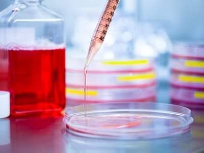
Viral Transport Media
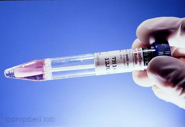
These chemicals are added as a way to “safely” transfer the patient sample to the lab for testing, culturing, and other molecular biological techniques. They often are composed of some sort of salt solution, fetal bovine serum, antibiotics, and can be contaminated by disinfectants such as ethanol. These are all substances which are toxic to cells and can change the sample before the culturing process even begins.
After this, they may do some centrifugation (spin the sample really fast) to separate out larger particles (leaving behind many EV’s, exosomes, “viruses,” etc. that are too small to be filtered out) and then will take what is called the supernatant, which is what is collected after letting the sample settle, and add it to a cell to be cultured.
There are many different types of cells that can be chosen from to culture a “virus” in and they typically come from either human cancer cells or from animals such as monkeys and rabbits:
“Examples of well-known cell types that are standard for most virology laboratories are primary rhesus monkey kidney (RhMK) cells, primary rabbit kidney cells, human lung fibroblasts (MRC-5), human foreskin fibroblasts, human epidermoid carcinoma cells (HEp-2), human lung carcinoma cells (A549), and others.”
https://www.ncbi.nlm.nih.gov/pmc/articles/PMC1797634/
The cells that are used are critical to producing a “virus” which makes choosing the right one extremely important. There are problems with this step which can lead to contamination, errors, and faulty research.
Cell Line Misidentification
There is a crisis in cell cultures which threatens to throw out the results of many studies. Cell lines have been continuously mislabeled or replaced by cells from different individuals, tissues, or species. This problem has been known since the 1960’s and instead of being corrected over the years, it has only grown worse. Over half of all studies using misidentified cell lines have come since the year 2000, well after the problem was discovered. The results of experiments from studies using misidentified cell lines are cited and built upon by other researchers confounding the problem and throwing uncertainty over the results over a vast amount of scientific literature.
2D Cell Culture System
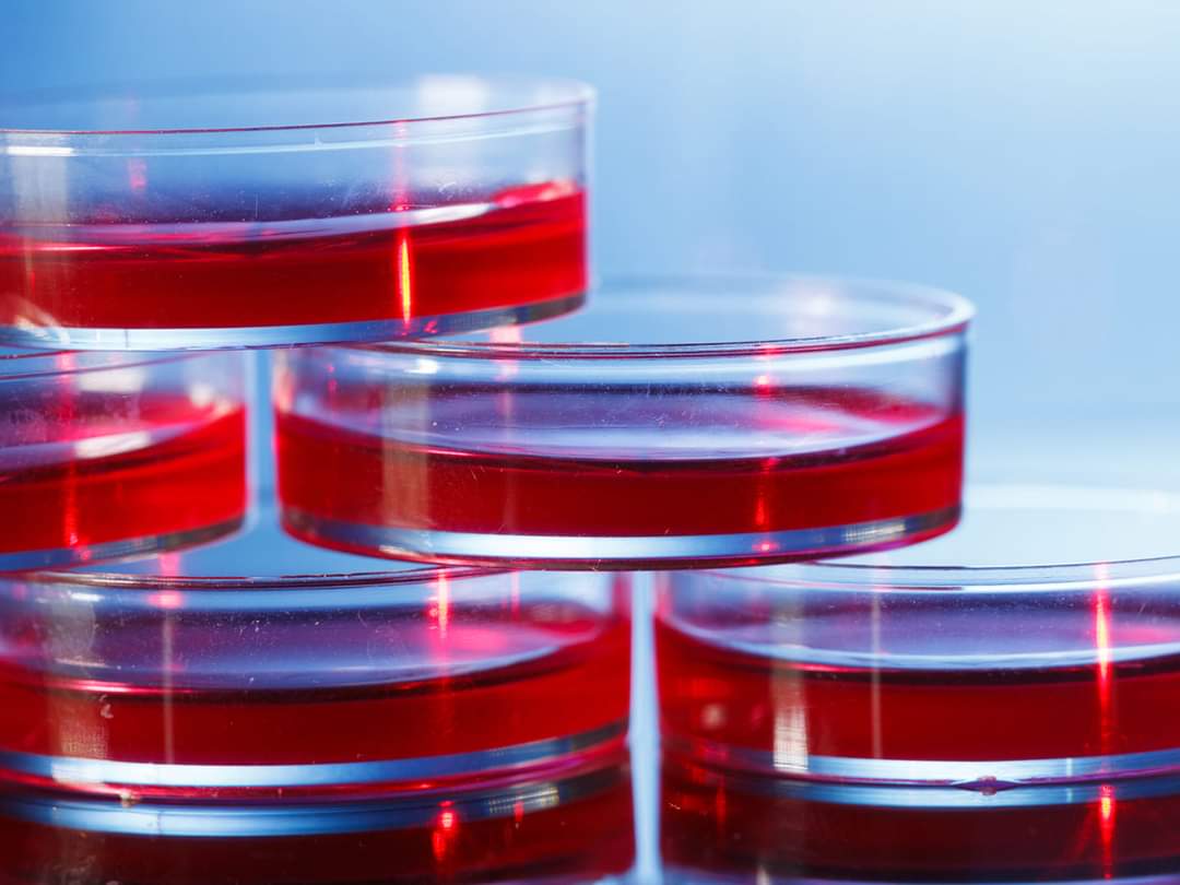
After the type of cell to be used is selected, the next step in the process is to choose the “environment” that the cells will be cultured in. For nearly all “virus” papers, the type of environment chosen is known as the 2D Cell Culture system. In this system, cells are grown on flat plastic petri dishes to which they then adhere to and spread. However, this system has many disadvantages such as the inability to mimic the in vivo (within a living organism) extracellular matrix microenvironment, the inability to predict results, and the inability to replicate the cellular microenvironment in terms of architecture, composition, physiological functions, and mechanical stimulus.
2D or not, 3D? That is the Question…
Cell Culture Media
Once the culture system is chosen, the cell is contained within cell culture media. There are many types that they choose from including both natural and artificial varieties. The actual composition and make-up of these media is unknown in most cases and can vary batch-to-batch but, like the VTM, they typically contain a salt solution, antibiotics, and fetal bovine serum as well as added amino acids, glucose, vitamins, and nutrients. The compounds that make up these media individually have been shown to be detrimental to cells and the combined effects are relatively unknown and understudied.
A quick look at a few of the compounds making up VTM and Cell Culture Media show how they can impact the results of the cell culture and why they should not be used.
Antibiotics
Antibiotics are regularly used in cell cultures in order to prevent bacterial contamination. However, it is well known that they are toxic to cells and their use impairs cell growth and differentiation. They have an effect on the metabolism of cultured cells, cell proliferation, differentiation, and gene expression. They damage mitochondria which results in genomic instability. They can also attack non-bacterial structures within the cell. There are many warnings against the use of antibiotics in cell culture as well as relying on data generated from them but these warnings are often ignored.
The Effects of Antibiotics on Cell Cultures
Fetal Bovine Serum
Fetal bovine serum is derived from the blood of the unborn fetus of slaughtered pregnant cows. It’s use is questionable not only on a moral ground as the fetus is normally alive as the blood is drained from its heart but also due to the fact that the RNA from the serum is nearly impossible to separate in the cell culture and can influence the results. It has also been shown to affect the genotypic and phenotypic response. The batches vary in composition and many of the components are unknown.
The Unethical use of Fetal Bovine Serum
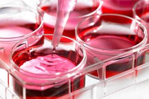
Once the cell and the cell culture media are chosen, the supernatant from the patient sample is added to the cell culture. Media/Antibiotics are added and changed throughout this process. An example from the Zhou study, one of the original “SARS-COV-2” papers:
“Cultured cell monolayers were maintained in their respective medium. The PCR-positive BALF sample from ICU-06 patient was spun at 8,000g for 15 min, filtered and diluted 1:2 with DMEM supplemented with 16 μg ml−1 trypsin before it was added to the cells. After incubation at 37 °C for 1 h, the inoculum was removed and replaced with fresh culture medium containing antibiotics (see below) AND 16 μg ml−1 trypsin. The cells were incubated at 37 °C and observed daily for cytopathogenic effects.”
https://www.nature.com/articles/s41586-020-2012-7
As can be seen from the above example, after the supernatant and the media are added, the cell culture is incubated and observed daily for what is called cytopathic or cytopathogenic effects.
The Cytopathic Effect
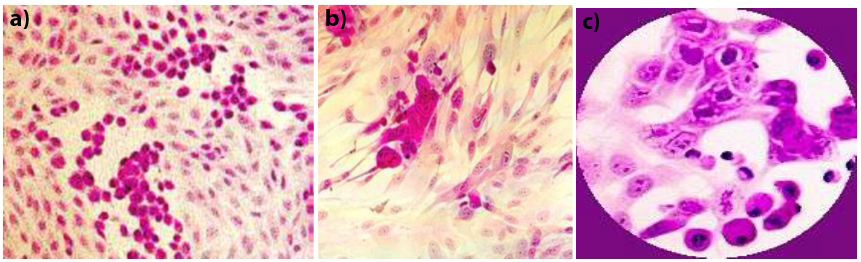
Cytopathic Effects are the holy grail of the cell culture experiment. These are structural changes to the host cell said to be caused by an invading “virus.” As “viruses” are unable to be seen without the use of an Electron Microscope, they look for this effect as INDIRECT evidence that a “virus” is present in the cell culture. If they observe CPE, they ASSUME a “virus” is present as this effect is supposedly specific to “viruses.” However, this is not the case at all as there are many other factors which can cause the very same effect such as: bacteria, parasites, amoebas, and chemical contaminants such as antibiotics and antifungals. CPE can also be due to the ageing of the cells, miscalculations in the growth media formula, and from the sub-culturing techniques utilized.
Creating the Cytopathic Effect
It’s clear to see that if CPE, the end result of a cell culture experiment used as proof of a “virus,” can be caused by other organisms and chemicals, CPE is not specific to “viruses.” Antibiotics and antifungals are pretty much a given to be in cell cultures at this point and alone are enough to cause CPE. However, it is also well known that these various other factors (bacteria, parasites, amoebas) are most likely in the culture as well.
Contamination
Cell culture contamination is not the exception but the rule. Virologists try to mitigate and control the effects of contamination yet admit there is no way that they can eliminate it. Contamination can come in many forms such as bacteria like mycoplasmas, other “viruses,” parasites, yeast, fungus, etc.
Cell Line Misidentification and Contamination
The Unwanted: Cell Culture Contaminants
Contamination can also come from the environment in various ways such as plastic ware chemicals leaching into the cell culture from petri dishes, organic/inorganic compounds in the water used invading the culture, and even effects from the types of lights used altering the cells. There are other factors such as temperature, atmospheric conditions, and pH levels to consider.
Sub-Culturing and Cell Culture Adaptation
Along with the numerous other sources of cell culture contamination that alter the cell is the issue of sub-culturing (or passaging) the cell culture before and during the culture experiment. Sub-culturing is the process of dividing and transferring cells of one culture that has outgrown the dish and/or used up its “nutrients” into another vessel with new growth media. The stress that this can cause to cells is known to have an impact on the cell morphology, gene expression, stimulus response, growth rate, etc. The cell culture adaptations can cause entirely new gene sequences to occur that were previously undetected.
Sub-Culturing and Cell Culture Adaptations
Passage in Vero Cells: The Variant Game
Reproducibility Crisis

Taking into consideration the numerous sources of contamination, the huge problem of misidentification of cell lines, the various chemicals/antibiotics/serum/animal cells etc. used in the cultures and the toxic, cell-altering effects they have, is it any wonder why there is a reproducibility crisis in science, especially regarding cell cultures? Experiments are rarely able to be reproduced which leads to the over 3 million “variants” of “SARS-COV-2” we are currently in the midst of seeing with a new one seemingly popping up every day.
Cell Culture Reproducibility Crisis
Once the supposed “viral” CPE is observed in the toxic cell culture, the unpurified supernatant is once again collected for EM imaging, genome sequencing, and animal testing. It should be clear from the various reasons listed above why this is not adequate proof of any “virus.”
- Without proper purification/isolation, there is absolutely no way to tell that the particle they pick out to be the representation of their “novel virus” in the EM image actually is a “virus” at all.
- Without purification and due to the numerous toxic ingredients added to the original sample, there is no way to confirm that the RNA/DNA used for the genome actually comes from one unaltered source.
- Without purification/isolation, there is no way to definitively say that there was a “virus” contained within the cell culture soup which is unnaturally shoved intranasally down the noses of test animals. If the animals do get sick, it could be due to the antibiotics, the FBS, the media, the nutrients, the contaminants, the stress of the experiments, or a combination of any of these factors.
It should be obvious that the end result of a cell culture experiment in no way reflects what was originally taken from the patient sample. The results in no way reflect reality. Cell cultures are nothing more than a recipe to create cell death which is blamed on invisible “viruses” never proven to exist.
For this, we locked down, quarantined, social-distanced, shut down economies and small businesses, shunned the elderly, masked, and vaccinated with rushed experimental gene therapies.
All based on “scientific” fraud.

Very clearly said, nice summary! Do you want me to point out typos or not? Don’t want to be a grammar Nazi… And your posts rarely have typos, amazingly! Another clue that you might not actually be human. ..lol…
LikeLiked by 1 person
YES! Please do! I hate typos and I always want to correct them.
LikeLike
I went through and found/corrected 2 or 3 typos. Do you think that was how much typos I had or were there more? I hate missing typos. 😦
LikeLike
“this system has many disadbantages such as the inability”
You might want to try grammarly.com, or if you want, I could run your articles through it for a grammar check.
LikeLiked by 1 person
Thank you for pointing out the typo and the suggestion! I appreciate it! I will give grammarly.com a shot. I try very hard to catch typos but they have a way of slipping by me from time to time. Thanks again for the help. 🙂
LikeLike
Any scientist performing science according to the scientific method would implement controls to account for the variables involved. For instance run the experiment without the lung fluid sample. Then run it with a fluid sample from the same person without antibiotics. Further – They should be reviewing the EM after the centrifugal process for the specific density where a virus would band… moreover what errors might be introduced in looking at the EM results from bombarding the specimen with a high amount of electrons… your detail bringing down to laymans terms is very helpful and insightful to understanding these at best inadequate procedures or at worst deliberate misleading procedures to arrive at a desired solution. Many thanks
LikeLiked by 1 person
Thanks, I appreciate the kind words and feedback 🙂 You are spot on with the lack of proper controls. This is such a glaring and huge problem that is seemingly swept under the rug. I’m glad that people like Dr. Lanka are bringing this issue to light.
LikeLike
Mike from what I have read, most viral cultures if not all only produce “a virus” when monkey vero kidney cells are used. While they may use the other cell types you mention, only fake virus grows on Vero cells. Can we please fact check this because I think I am right.
Great blog. I’ve referred this to Tom Cowan and he’s already turned people on to your blog. You will be getting a lot of traffic. Well done.
LikeLiked by 1 person
Thanks for the kind words and support! I really appreciate it. 🙂
While it seems that Vero cells are the go to cell line these days for “viral” culture, they do use other cell lines as well such as MDCK, A549, HEK293, etc. Here is a table with some information on the cell used as well as the CPE caused by different cell lines for different “viruses:”
https://www.ncbi.nlm.nih.gov/labs/pmc/articles/PMC1797634/table/t2/?report=objectonly
LikeLike
Regarding the recent discovery of (ahem!) vaccine derived polio virus in sewage from London, I decided to research how alleged viruses are detected in sewage. I found the following link; https://file.scirp.org/pdf/AiM_2013091110391866.pdf in which they talk about “plaque assays” allegedly a “gold standard”! https://www.ncbi.nlm.nih.gov/pmc/articles/PMC7300432/#cpmc105-bib-0015 It seems to me that “plaque assays” are another name for cell culture, am I missing something?
LikeLike
Reblogged this on Gustirna.
LikeLike